

40
M I C H A E L L O C K E
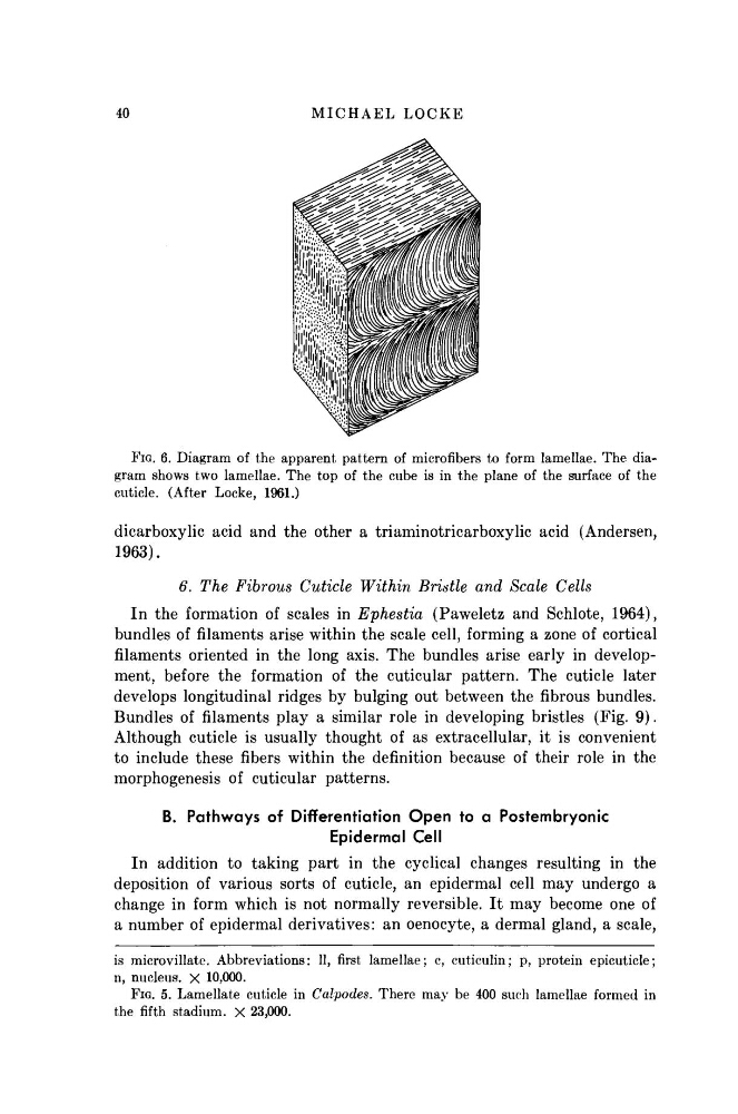
FIG. 6. Diagram of the apparent pattern of micro fibers to form lamellae. The diagram shows two lamellae. The top of the cube is in the plane of the surface of the
cuticle. (After Locke, 1961.)
dicarboxylic acid and the other a triaminotricarboxylic acid (Andersen,
1963).
6. The Fibrous Cuticle Within Bristle and Scale Cells
In the formation of scales in Ephestia (Paweletz and Schlote, 1964),
bundles of filaments arise within the scale cell, forming a zone of cortical
filaments oriented in the long axis. The bundles arise early in development, before the formation of the cuticular pattern. The cuticle later
develops longitudinal ridges by bulging out between the fibrous bundles.
Bundles of filaments play a similar role in developing bristles (Fig. 9).
Although cuticle is usually thought of as extracellular, it is convenient
to include these fibers within the definition because of their role in the
morphogenesis of cuticular patterns.
B. Pathways of Differentiation Open to a Postembryonic
Epidermal Cell
In addition to taking part in the cyclical changes resulting in the
deposition of various sorts of cuticle, an epidermal cell may undergo a
change in form which is not normally reversible. It may become one of
a number of epidermal derivatives: an oenocyte, a dermal gland, a scale,
is microvillate. Abbreviations: 11, first lamellae; c, cuticulin; p, protein epicuticle;
n, nucleus. X 10,000.
FIG. 5. Lamellate cuticle in Calpodes. There may be 400 such lamellae formed in
the fifth stadium. X 23,000.
M I C H A E L L O C K E
FIG. 6. Diagram of the apparent pattern of micro fibers to form lamellae. The diagram shows two lamellae. The top of the cube is in the plane of the surface of the
cuticle. (After Locke, 1961.)
dicarboxylic acid and the other a triaminotricarboxylic acid (Andersen,
1963).
6. The Fibrous Cuticle Within Bristle and Scale Cells
In the formation of scales in Ephestia (Paweletz and Schlote, 1964),
bundles of filaments arise within the scale cell, forming a zone of cortical
filaments oriented in the long axis. The bundles arise early in development, before the formation of the cuticular pattern. The cuticle later
develops longitudinal ridges by bulging out between the fibrous bundles.
Bundles of filaments play a similar role in developing bristles (Fig. 9).
Although cuticle is usually thought of as extracellular, it is convenient
to include these fibers within the definition because of their role in the
morphogenesis of cuticular patterns.
B. Pathways of Differentiation Open to a Postembryonic
Epidermal Cell
In addition to taking part in the cyclical changes resulting in the
deposition of various sorts of cuticle, an epidermal cell may undergo a
change in form which is not normally reversible. It may become one of
a number of epidermal derivatives: an oenocyte, a dermal gland, a scale,
is microvillate. Abbreviations: 11, first lamellae; c, cuticulin; p, protein epicuticle;
n, nucleus. X 10,000.
FIG. 5. Lamellate cuticle in Calpodes. There may be 400 such lamellae formed in
the fifth stadium. X 23,000.