

I . P R O B L E M
O F T H E
O R G A N I Z E R
43
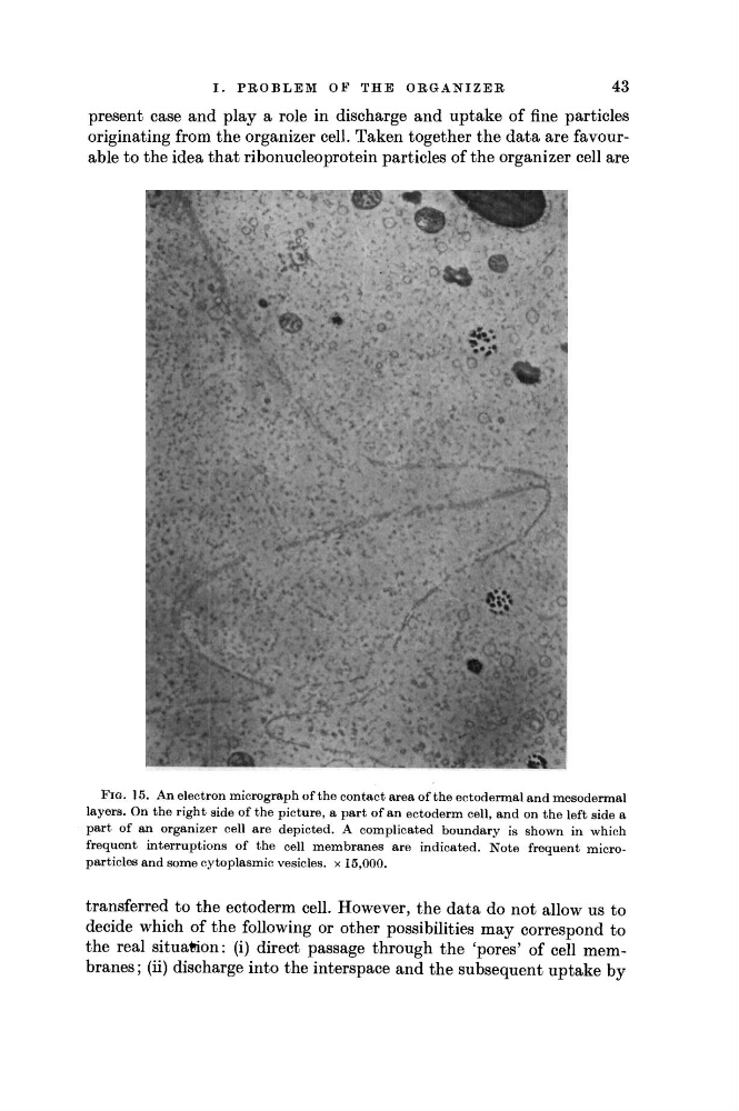
present case and play a role in discharge and uptake of fine particles
originating from the organizer cell. Taken together the data are favourable to the idea that ribonucleoprotein particles of the organizer cell are
F I G . 1 5 . A n electron micrograph of the contact area of the ectodermal and mesodermal
layers. On the right side of the picture, a part of an ectoderm cell, and on the left side a
part of an organizer cell are depicted. A complicated boundary is shown in which
frequent interruptions of the cell membranes are indicated. N o t e frequent microparticles and some cytoplasmic vesicles, χ 1 5 , 0 0 0 .
transferred to the ectoderm cell. However, the data do not allow us to
decide which of the following or other possibilities may correspond to
the real situation: (i) direct passage through the 'pores' of cell membranes ; (ii) discharge into the interspace and the subsequent uptake by
O F T H E
O R G A N I Z E R
43
present case and play a role in discharge and uptake of fine particles
originating from the organizer cell. Taken together the data are favourable to the idea that ribonucleoprotein particles of the organizer cell are
F I G . 1 5 . A n electron micrograph of the contact area of the ectodermal and mesodermal
layers. On the right side of the picture, a part of an ectoderm cell, and on the left side a
part of an organizer cell are depicted. A complicated boundary is shown in which
frequent interruptions of the cell membranes are indicated. N o t e frequent microparticles and some cytoplasmic vesicles, χ 1 5 , 0 0 0 .
transferred to the ectoderm cell. However, the data do not allow us to
decide which of the following or other possibilities may correspond to
the real situation: (i) direct passage through the 'pores' of cell membranes ; (ii) discharge into the interspace and the subsequent uptake by