

190
F.
C.
STEWARD
A N D
Η.
Y.
MOHAN
RAM
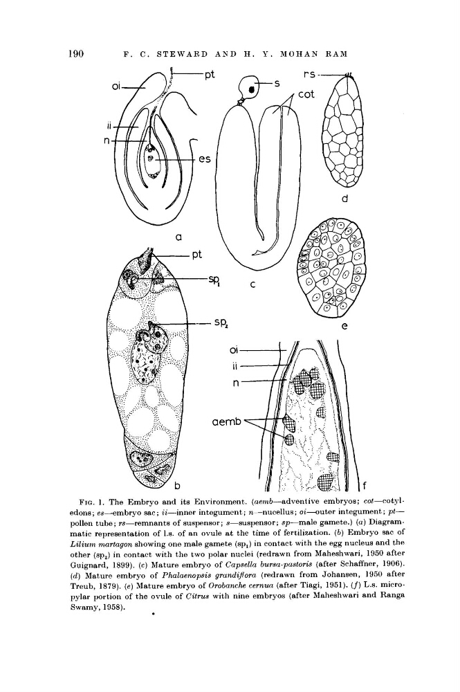
FIG. 1. The Embryo and its Environment, (aemb—adventive
embryos;
cot—cotyledons ; es—embryo sac; it—inner integument; η—nucellus;
οί—outer integument; pt—
pollen tube; rs—remnants
of suspensor; s—suspensor;
sp—male gamete.) (a) Diagrammatic representation of I.e. of an ovule at the time of fertilization, (b) Embryo sac of
Lilium martagón showing one male gamete (sp x ) in contact with the egg nucleus and the
other (sp 2 ) in contact with the two polar nuclei (redrawn from Maheshwari, 1950 after
Guignard, 1899). (c) Mature embryo of Capsula bursa-pastoris (after Schaffner, 1906).
(d) Mature embryo of Phalaenopsis
grandiflora (redrawn from Johansen, 1950 after
Treub, 1879). (e) Mature embryo of Orobanche cernua (after Tiagi, 1951). (/) L.s. micropylar portion of the ovule of Citrus with nine embryos (after Maheshwari and Ranga
Swamy, 1958).
F.
C.
STEWARD
A N D
Η.
Y.
MOHAN
RAM
FIG. 1. The Embryo and its Environment, (aemb—adventive
embryos;
cot—cotyledons ; es—embryo sac; it—inner integument; η—nucellus;
οί—outer integument; pt—
pollen tube; rs—remnants
of suspensor; s—suspensor;
sp—male gamete.) (a) Diagrammatic representation of I.e. of an ovule at the time of fertilization, (b) Embryo sac of
Lilium martagón showing one male gamete (sp x ) in contact with the egg nucleus and the
other (sp 2 ) in contact with the two polar nuclei (redrawn from Maheshwari, 1950 after
Guignard, 1899). (c) Mature embryo of Capsula bursa-pastoris (after Schaffner, 1906).
(d) Mature embryo of Phalaenopsis
grandiflora (redrawn from Johansen, 1950 after
Treub, 1879). (e) Mature embryo of Orobanche cernua (after Tiagi, 1951). (/) L.s. micropylar portion of the ovule of Citrus with nine embryos (after Maheshwari and Ranga
Swamy, 1958).