

30
J A C K C O H E N
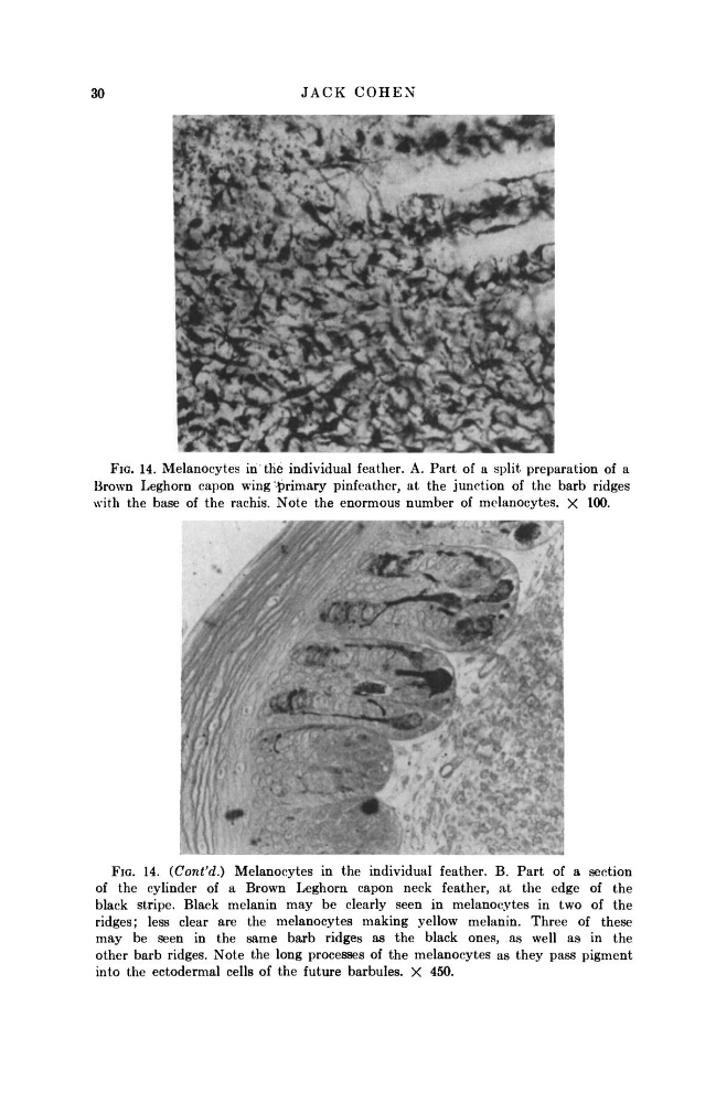
FIG. 14. Melanocytes in tile individual feather. A. Part of a split preparation of a
Brown Leghorn capon wing primary pinfeather, at the junction of the barb ridges
with the base of the rachis. Note the enormous number of melanocytes. X 100.
FIG. 14. (Cont'd.) Melanocytes in the individual feather. B. Part of a section
of the cylinder of a Brown Leghorn capon neck feather, at the edge of the
black stripe. Black melanin may be clearly seen in melanocytes in two of the
ridges; less clear are the melanocytes making yellow melanin. Three of these
may be seen in the same barb ridges as the black ones, as well as in the
other barb ridges. Note the long processes of the melanocytes as they pass pigment
into the ectodermal cells of the future barbules. X 450.
J A C K C O H E N
FIG. 14. Melanocytes in tile individual feather. A. Part of a split preparation of a
Brown Leghorn capon wing primary pinfeather, at the junction of the barb ridges
with the base of the rachis. Note the enormous number of melanocytes. X 100.
FIG. 14. (Cont'd.) Melanocytes in the individual feather. B. Part of a section
of the cylinder of a Brown Leghorn capon neck feather, at the edge of the
black stripe. Black melanin may be clearly seen in melanocytes in two of the
ridges; less clear are the melanocytes making yellow melanin. Three of these
may be seen in the same barb ridges as the black ones, as well as in the
other barb ridges. Note the long processes of the melanocytes as they pass pigment
into the ectodermal cells of the future barbules. X 450.