

298
JOHN RUNNSTRÖM
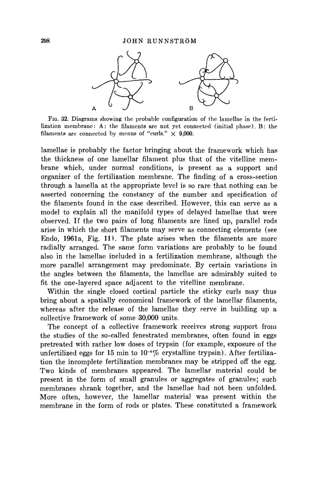
FIG. 32. Diagrams showing the probable configuration of the lamellae in the fertilization membrane: A: the filaments are not yet connected (initial phase). B: the
filaments are connected by means of "curls." X 9,000.
lamellae is probably the factor bringing about the framework which has
the thickness of one lamellar filament plus that of the vitelline membrane which, under normal conditions, is present as a support and
organizer of the fertilization membrane. The rinding of a cross-section
through a lamella at the appropriate level is so rare that nothing can be
asserted concerning the constancy of the number and specification of
the filaments found in the case described. However, this can serve as a
model to explain all the manifold types of delayed lamellae that were
observed. If the two pairs of long filaments are lined up, parallel rods
arise in which the short filaments may serve as connecting elements (see
Endo, 1961a, Fig. 11). The plate arises when the filaments are more
radially arranged. The same form variations are probably to be found
also in the lamellae included in a fertilization membrane, although the
more parallel arrangement may predominate. By certain variations in
the angles between the filaments, the lamellae are admirably suited to
fit the one-layered space adjacent to the vitelline membrane.
Within the single closed cortical particle the sticky curls may thus
bring about a spatially economical framework of the lamellar filaments,
whereas after the release of the lamellae they serve in building up a
collective framework of some 30,000 units.
The concept of a collective framework receives strong support from
the studies of the so-called fenestrated membranes, often found in eggs
pretreated with rather low doses of trypsin (for example, exposure of the
unfertilized eggs for 15 min to 10"
4
% crystalline trypsin). After fertilization the incomplete fertilization membranes may be stripped off the egg.
Two kinds of membranes appeared. The lamellar material could be
present in the form of small granules or aggregates of granules; such
membranes shrank together, and the lamellae had not been unfolded.
More often, however, the lamellar material was present within the
membrane in the form of rods or plates. These constituted a framework
JOHN RUNNSTRÖM
FIG. 32. Diagrams showing the probable configuration of the lamellae in the fertilization membrane: A: the filaments are not yet connected (initial phase). B: the
filaments are connected by means of "curls." X 9,000.
lamellae is probably the factor bringing about the framework which has
the thickness of one lamellar filament plus that of the vitelline membrane which, under normal conditions, is present as a support and
organizer of the fertilization membrane. The rinding of a cross-section
through a lamella at the appropriate level is so rare that nothing can be
asserted concerning the constancy of the number and specification of
the filaments found in the case described. However, this can serve as a
model to explain all the manifold types of delayed lamellae that were
observed. If the two pairs of long filaments are lined up, parallel rods
arise in which the short filaments may serve as connecting elements (see
Endo, 1961a, Fig. 11). The plate arises when the filaments are more
radially arranged. The same form variations are probably to be found
also in the lamellae included in a fertilization membrane, although the
more parallel arrangement may predominate. By certain variations in
the angles between the filaments, the lamellae are admirably suited to
fit the one-layered space adjacent to the vitelline membrane.
Within the single closed cortical particle the sticky curls may thus
bring about a spatially economical framework of the lamellar filaments,
whereas after the release of the lamellae they serve in building up a
collective framework of some 30,000 units.
The concept of a collective framework receives strong support from
the studies of the so-called fenestrated membranes, often found in eggs
pretreated with rather low doses of trypsin (for example, exposure of the
unfertilized eggs for 15 min to 10"
4
% crystalline trypsin). After fertilization the incomplete fertilization membranes may be stripped off the egg.
Two kinds of membranes appeared. The lamellar material could be
present in the form of small granules or aggregates of granules; such
membranes shrank together, and the lamellae had not been unfolded.
More often, however, the lamellar material was present within the
membrane in the form of rods or plates. These constituted a framework