

2.000
1.500
1000
0.500
230 240 250 260 270 280 290 300 310 320
πψ
2.000
1.500
1.000
0.500
(Β)
; vin
230 240 250 260 270 280 290 300
310 320
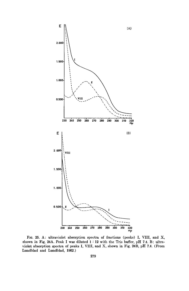
FIG. 25. A: ultraviolet absorption spectra of fractions (peaks) I, VIII, and X,
shown in Fig. 24A. Peak I was diluted 1 : 12 with the Tris buffer, pH 7.4. B: ultraviolet absorption spectra of peaks I, VIII, and X, shown in Fig. 24B, pH 7.4. (From
Lundblad and Lundblad, 1962.)
279
1.500
1000
0.500
230 240 250 260 270 280 290 300 310 320
πψ
2.000
1.500
1.000
0.500
(Β)
; vin
230 240 250 260 270 280 290 300
310 320
FIG. 25. A: ultraviolet absorption spectra of fractions (peaks) I, VIII, and X,
shown in Fig. 24A. Peak I was diluted 1 : 12 with the Tris buffer, pH 7.4. B: ultraviolet absorption spectra of peaks I, VIII, and X, shown in Fig. 24B, pH 7.4. (From
Lundblad and Lundblad, 1962.)
279