

VITELLINE MEMBRANE AND CORTICAL PARTICLES 277
(A)
1
10
20
30
AO
50
60
70
80
90
100 110
Γ
J
l
Τ
Î Fraction No
1
10
20
»
40
50
60
70
80
90
100 110 120
Î
Τ
Τ
Τ
Τ Fraction No
12
3
A
5
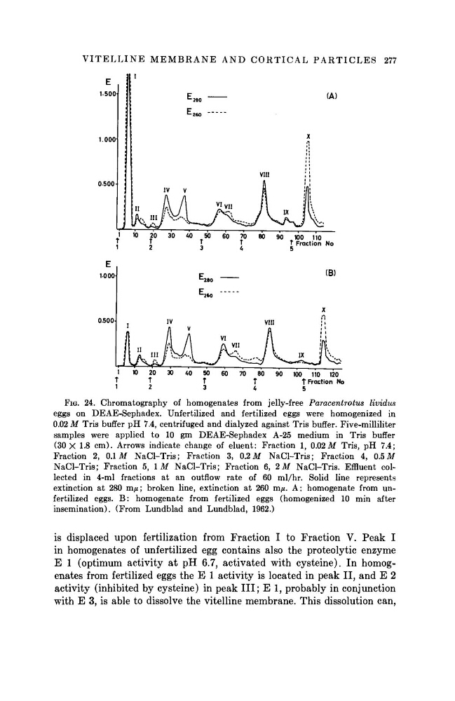
FIG. 24. Chromatography of homogenates from jelly-free Paracentrotus
lividus
eggs on DEAE-Sephadex. Unfertilized and fertilized eggs were homogenized in
0.02 M Tris buffer pH 7.4, centrifuged and dialyzed against Tris buffer. Five-milliliter
samples were applied to 10 gm DEAE-Sephadex A-25 medium in Tris buffer
(30 X 1.8 cm). Arrows indicate change of eluent: Fraction 1, 0.02 M Tris, pH 7.4;
Fraction 2, 0.1 M NaCl-Tris; Fraction 3, 0.2 M NaCl-Tris; Fraction 4, 0.5 M
NaCl-Tris; Fraction 5, 1M NaCl-Tris; Fraction 6, 2 M NaCl-Tris. Effluent collected in 4-ml fractions at an outflow rate of 60 ml/hr. Solid line represents
extinction at 280 τημ; broken line, extinction at 260 m/*. A: homogenate from unfertilized eggs. B: homogenate from fertilized eggs (homogenized 10 min after
insemination). (From Lundblad and Lundblad, 1962.)
is displaced upon fertilization from Fraction I to Fraction V. Peak I
in homogenates of unfertilized egg contains also the proteolytic enzyme
Ε 1 (optimum activity at pH 6.7, activated with cysteine). In homogenates from fertilized eggs the Ε 1 activity is located in peak II, and Ε 2
activity (inhibited by cysteine) in peak III; Ε 1, probably in conjunction
with Ε 3, is able to dissolve the vitelline membrane. This dissolution can,
(A)
1
10
20
30
AO
50
60
70
80
90
100 110
Γ
J
l
Τ
Î Fraction No
1
10
20
»
40
50
60
70
80
90
100 110 120
Î
Τ
Τ
Τ
Τ Fraction No
12
3
A
5
FIG. 24. Chromatography of homogenates from jelly-free Paracentrotus
lividus
eggs on DEAE-Sephadex. Unfertilized and fertilized eggs were homogenized in
0.02 M Tris buffer pH 7.4, centrifuged and dialyzed against Tris buffer. Five-milliliter
samples were applied to 10 gm DEAE-Sephadex A-25 medium in Tris buffer
(30 X 1.8 cm). Arrows indicate change of eluent: Fraction 1, 0.02 M Tris, pH 7.4;
Fraction 2, 0.1 M NaCl-Tris; Fraction 3, 0.2 M NaCl-Tris; Fraction 4, 0.5 M
NaCl-Tris; Fraction 5, 1M NaCl-Tris; Fraction 6, 2 M NaCl-Tris. Effluent collected in 4-ml fractions at an outflow rate of 60 ml/hr. Solid line represents
extinction at 280 τημ; broken line, extinction at 260 m/*. A: homogenate from unfertilized eggs. B: homogenate from fertilized eggs (homogenized 10 min after
insemination). (From Lundblad and Lundblad, 1962.)
is displaced upon fertilization from Fraction I to Fraction V. Peak I
in homogenates of unfertilized egg contains also the proteolytic enzyme
Ε 1 (optimum activity at pH 6.7, activated with cysteine). In homogenates from fertilized eggs the Ε 1 activity is located in peak II, and Ε 2
activity (inhibited by cysteine) in peak III; Ε 1, probably in conjunction
with Ε 3, is able to dissolve the vitelline membrane. This dissolution can,