

244
J O H N
R U N N S T R Ö M
in the elongated stalk by which the oocyte is fixed to the ovarian wall.
This stalk m a y be very electron dense. At a certain stage, numerous
vacuoles with variable diameter up to about 0.12 μ are present. I t is our
impression t h a t these vacuoles fuse to form lacunae in which the
cortical particles develop. At the same time, however, dense material is
budding, so as to be surrounded by the liquid space of the lacuna. The
dense material is the germ of the lamella and the extralamellar bodies.
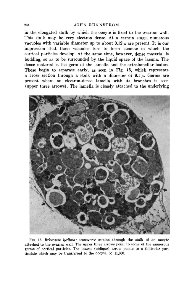
These begin to separate early, as seen in Fig. 15, which represents
a cross section through a stalk with a diameter of 9.1 μ. Germs are
present where an electron-dense lamella with its branches is seen
(upper three arrows). The lamella is closely attached to the underlying
FIG. 15. Brissopsis lynfera: transverse section through the stalk of an oocyte
attached to the ovarian wall. The upper three arrows point to some of the numerous
germs of cortical particles. The lowest (oblique) arrow points to a follicular particulate which may be transferred to the oocyte. X 11,000.
J O H N
R U N N S T R Ö M
in the elongated stalk by which the oocyte is fixed to the ovarian wall.
This stalk m a y be very electron dense. At a certain stage, numerous
vacuoles with variable diameter up to about 0.12 μ are present. I t is our
impression t h a t these vacuoles fuse to form lacunae in which the
cortical particles develop. At the same time, however, dense material is
budding, so as to be surrounded by the liquid space of the lacuna. The
dense material is the germ of the lamella and the extralamellar bodies.
These begin to separate early, as seen in Fig. 15, which represents
a cross section through a stalk with a diameter of 9.1 μ. Germs are
present where an electron-dense lamella with its branches is seen
(upper three arrows). The lamella is closely attached to the underlying
FIG. 15. Brissopsis lynfera: transverse section through the stalk of an oocyte
attached to the ovarian wall. The upper three arrows point to some of the numerous
germs of cortical particles. The lowest (oblique) arrow points to a follicular particulate which may be transferred to the oocyte. X 11,000.