

V I T E L L I N E M E M B R A N E A N D C O R T I C A L P A R T I C L E S 231
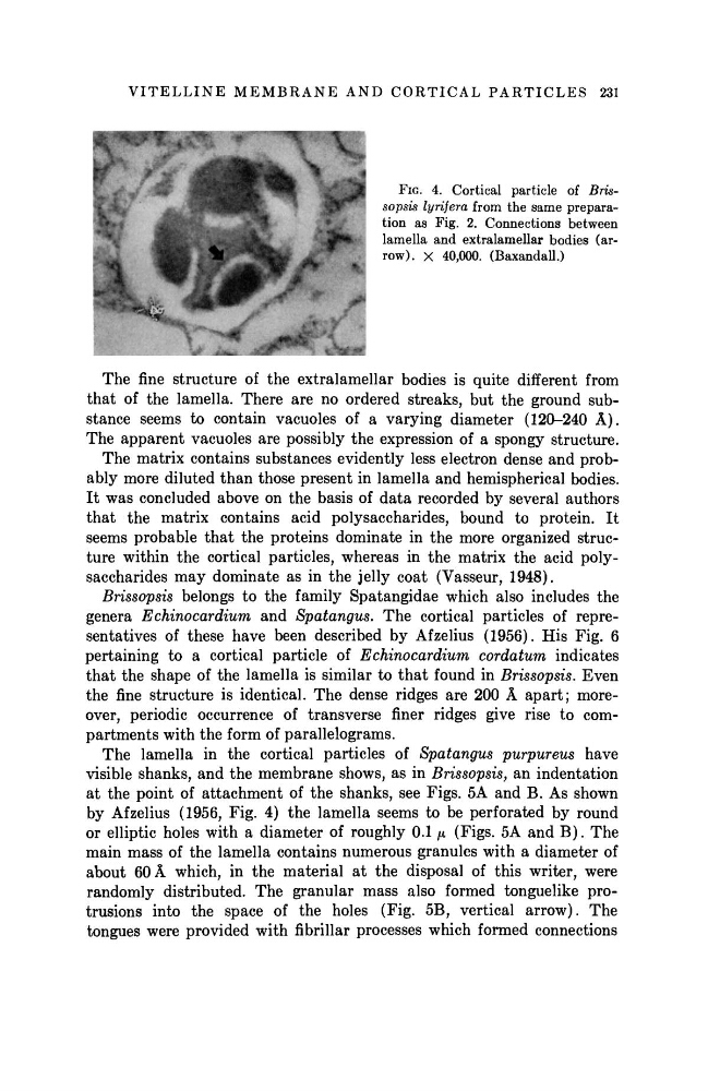
FIG. 4. Cortical particle of Brissopsis lyrifera from the same preparation as Fig. 2. Connections between
lamella and extralamellar bodies (arrow), χ 40,000. (Baxandall.)
The fine structure of the extralamellar bodies is quite different from
t h a t of the lamella. There are no ordered streaks, but the ground substance seems to contain vacuoles of a varying diameter (120-240 Â ) .
The apparent vacuoles are possibly the expression of a spongy structure.
The matrix contains substances evidently less electron dense and probably more diluted t h a n those present in lamella and hemispherical bodies.
I t was concluded above on the basis of d a t a recorded by several authors
t h a t the matrix contains acid polysaccharides, bound to protein. It
seems probable t h a t the proteins dominate in the more organized structure within the cortical particles, whereas in the matrix the acid polysaccharides m a y dominate as in the jelly coat (Vasseur, 1948).
Brissopsis belongs to the family Spatangidae which also includes the
genera Echinocardium
and Spatangus.
The cortical particles of representatives of these have been described by Afzelius (1956). His Fig. 6
pertaining to a cortical particle of Echinocardium
cordatum
indicates
t h a t the shape of the lamella is similar to t h a t found in Brissopsis. Even
the fine structure is identical. The dense ridges are 200 Â a p a r t ; moreover, periodic occurrence of transverse finer ridges give rise to compartments with the form of parallelograms.
The lamella in the cortical particles of Spatangus
purpureus
have
visible shanks, and the membrane shows, as in Brissopsis, an indentation
at the point of attachment of the shanks, see Figs. 5A and B . As shown
by Afzelius (1956, Fig. 4) the lamella seems to be perforated by round
or elliptic holes with a diameter of roughly 0.1 μ (Figs. 5A and B ) . The
main mass of the lamella contains numerous granules with a diameter of
about 60 Â which, in the material at the disposal of this writer, were
randomly distributed. The granular mass also formed tonguelike protrusions into the space of the holes (Fig. 5B, vertical arrow). The
tongues were provided with fibrillar processes which formed connections
FIG. 4. Cortical particle of Brissopsis lyrifera from the same preparation as Fig. 2. Connections between
lamella and extralamellar bodies (arrow), χ 40,000. (Baxandall.)
The fine structure of the extralamellar bodies is quite different from
t h a t of the lamella. There are no ordered streaks, but the ground substance seems to contain vacuoles of a varying diameter (120-240 Â ) .
The apparent vacuoles are possibly the expression of a spongy structure.
The matrix contains substances evidently less electron dense and probably more diluted t h a n those present in lamella and hemispherical bodies.
I t was concluded above on the basis of d a t a recorded by several authors
t h a t the matrix contains acid polysaccharides, bound to protein. It
seems probable t h a t the proteins dominate in the more organized structure within the cortical particles, whereas in the matrix the acid polysaccharides m a y dominate as in the jelly coat (Vasseur, 1948).
Brissopsis belongs to the family Spatangidae which also includes the
genera Echinocardium
and Spatangus.
The cortical particles of representatives of these have been described by Afzelius (1956). His Fig. 6
pertaining to a cortical particle of Echinocardium
cordatum
indicates
t h a t the shape of the lamella is similar to t h a t found in Brissopsis. Even
the fine structure is identical. The dense ridges are 200 Â a p a r t ; moreover, periodic occurrence of transverse finer ridges give rise to compartments with the form of parallelograms.
The lamella in the cortical particles of Spatangus
purpureus
have
visible shanks, and the membrane shows, as in Brissopsis, an indentation
at the point of attachment of the shanks, see Figs. 5A and B . As shown
by Afzelius (1956, Fig. 4) the lamella seems to be perforated by round
or elliptic holes with a diameter of roughly 0.1 μ (Figs. 5A and B ) . The
main mass of the lamella contains numerous granules with a diameter of
about 60 Â which, in the material at the disposal of this writer, were
randomly distributed. The granular mass also formed tonguelike protrusions into the space of the holes (Fig. 5B, vertical arrow). The
tongues were provided with fibrillar processes which formed connections