

DETERMINATION OF NEURAL CONNECTIONS
187
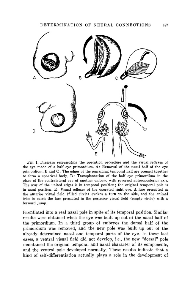
FIG. 1. Diagram representing the operation procedure and the visual reflexes of
the eye made of a half eye primordium. A: Removal of the nasal half of the eye
primordium. Β and C: The edges of the remaining temporal half are pressed together
to form a spherical body. D: Transplantation of the half eye primordium in the
place of the contralateral eye of another embryo with reversed anteroposterior axis.
The scar of the united edges is in temporal position; the original temporal pole is
in nasal position. E: Visual reflexes of the operated right eye. A lure presented in
the anterior visual field (filled circle) evokes a turn to the side, and the animal
tries to catch the lure presented in the posterior visual field (empty circle) with a
forward jump.
ferentiated into a real nasal pole in spite of its temporal position. Similar
results were obtained when the eye was built up out of the nasal half of
the primordium. In a third group of embryos the dorsal half of the
primordium was removed, and the new pole was built up out of the
already determined nasal and temporal parts of the eye. In these last
cases, a ventral visual field did not develop, i.e., the new "dorsal" pole
maintained the original temporal and nasal character of its components,
and the ventral pole developed normally. These results indicate that a
kind of self-differentiation actually plays a role in the development of
187
FIG. 1. Diagram representing the operation procedure and the visual reflexes of
the eye made of a half eye primordium. A: Removal of the nasal half of the eye
primordium. Β and C: The edges of the remaining temporal half are pressed together
to form a spherical body. D: Transplantation of the half eye primordium in the
place of the contralateral eye of another embryo with reversed anteroposterior axis.
The scar of the united edges is in temporal position; the original temporal pole is
in nasal position. E: Visual reflexes of the operated right eye. A lure presented in
the anterior visual field (filled circle) evokes a turn to the side, and the animal
tries to catch the lure presented in the posterior visual field (empty circle) with a
forward jump.
ferentiated into a real nasal pole in spite of its temporal position. Similar
results were obtained when the eye was built up out of the nasal half of
the primordium. In a third group of embryos the dorsal half of the
primordium was removed, and the new pole was built up out of the
already determined nasal and temporal parts of the eye. In these last
cases, a ventral visual field did not develop, i.e., the new "dorsal" pole
maintained the original temporal and nasal character of its components,
and the ventral pole developed normally. These results indicate that a
kind of self-differentiation actually plays a role in the development of