

170
SAUL WISCHNITZER
one end of a mitochondrion, and they exhibit the characteristic crystalline
structure of a yolk platelet core.
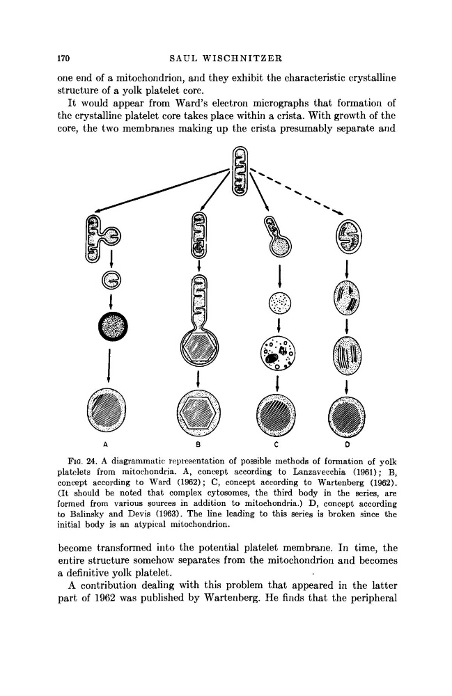
It would appear from Ward's electron micrographs that formation of
the crystalline platelet core takes place within a crista. With growth of the
core, the two membranes making up the crista presumably separate and
A
BCD
FIG. 24. A diagrammatic representation of possible methods of formation of yolk
platelets from mitochondria. A, concept according to Lanzavecchia (1961); B,
concept according to Ward (1962); C, concept according to Wartenberg (1962).
(It should be noted that complex cytosomes, the third body in the series, are
formed from various sources in addition to mitochondria.) D, concept according
to Balinsky and Devis (1963). The line leading to this series is broken since the
initial body is an atypical mitochondrion.
become transformed into the potential platelet membrane. In time, the
entire structure somehow separates from the mitochondrion and becomes
a definitive yolk platelet.
A contribution dealing with this problem that appeared in the latter
part of 1962 was published by Wartenberg. He finds that the peripheral
SAUL WISCHNITZER
one end of a mitochondrion, and they exhibit the characteristic crystalline
structure of a yolk platelet core.
It would appear from Ward's electron micrographs that formation of
the crystalline platelet core takes place within a crista. With growth of the
core, the two membranes making up the crista presumably separate and
A
BCD
FIG. 24. A diagrammatic representation of possible methods of formation of yolk
platelets from mitochondria. A, concept according to Lanzavecchia (1961); B,
concept according to Ward (1962); C, concept according to Wartenberg (1962).
(It should be noted that complex cytosomes, the third body in the series, are
formed from various sources in addition to mitochondria.) D, concept according
to Balinsky and Devis (1963). The line leading to this series is broken since the
initial body is an atypical mitochondrion.
become transformed into the potential platelet membrane. In time, the
entire structure somehow separates from the mitochondrion and becomes
a definitive yolk platelet.
A contribution dealing with this problem that appeared in the latter
part of 1962 was published by Wartenberg. He finds that the peripheral