

ULTRASTRUCTURE OF DEVELOPING AMPHIBIAN EGG
167
Kessel (1965), and Wischnitzer (1966) have clearly shown that ribosomes
are associated with the annulate lamellae. On the basis of this direct evidence as well as the fact that lamellae appear to be limited to very highly
metabolic cells and tissues, both normal and malignant, it is reasonable
to consider this organelle as an intermediate stage in the formation of
rough-surfaced cisternae. They would thus represent a structural precursor
of a form of endoplasmic reticulum.
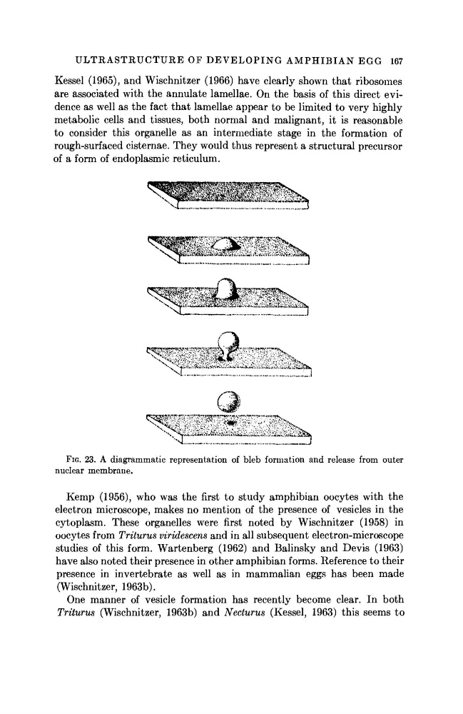
FIG. 23. A diagrammatic representation of bleb formation and release from outer
nuclear membrane.
Kemp (1956), who was the first to study amphibian oocytes with the
electron microscope, makes no mention of the presence of vesicles in the
cytoplasm. These organelles were first noted by Wischnitzer (1958) in
oocytes from Triturus viridescens and in all subsequent electron-microscope
studies of this form. Wartenberg (1962) and Balinsky and Devis (1963)
have also noted their presence in other amphibian forms. Reference to their
presence in invertebrate as well as in mammalian eggs has been made
(Wischnitzer, 1963b).
One manner of vesicle formation has recently become clear. In both
Triturus (Wischnitzer, 1963b) and Necturus (Kessel, 1963) this seems to
167
Kessel (1965), and Wischnitzer (1966) have clearly shown that ribosomes
are associated with the annulate lamellae. On the basis of this direct evidence as well as the fact that lamellae appear to be limited to very highly
metabolic cells and tissues, both normal and malignant, it is reasonable
to consider this organelle as an intermediate stage in the formation of
rough-surfaced cisternae. They would thus represent a structural precursor
of a form of endoplasmic reticulum.
FIG. 23. A diagrammatic representation of bleb formation and release from outer
nuclear membrane.
Kemp (1956), who was the first to study amphibian oocytes with the
electron microscope, makes no mention of the presence of vesicles in the
cytoplasm. These organelles were first noted by Wischnitzer (1958) in
oocytes from Triturus viridescens and in all subsequent electron-microscope
studies of this form. Wartenberg (1962) and Balinsky and Devis (1963)
have also noted their presence in other amphibian forms. Reference to their
presence in invertebrate as well as in mammalian eggs has been made
(Wischnitzer, 1963b).
One manner of vesicle formation has recently become clear. In both
Triturus (Wischnitzer, 1963b) and Necturus (Kessel, 1963) this seems to