

146
SAUL WISCHNITZER
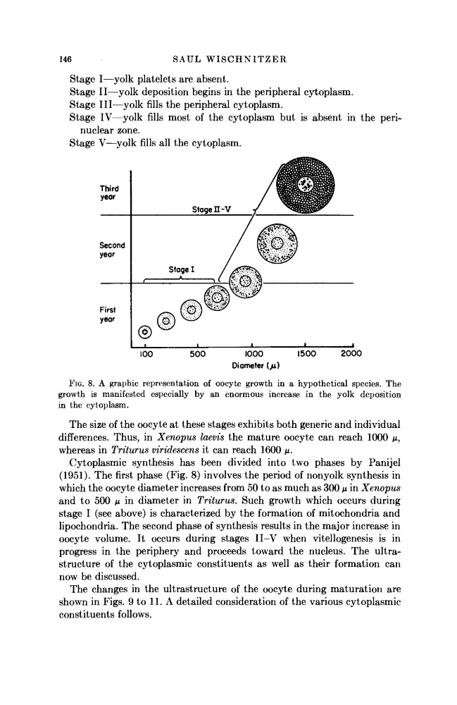
Stage I—yolk platelets are absent.
Stage II—yolk deposition begins in the peripheral cytoplasm.
Stage III—yolk fills the peripheral cytoplasm.
Stage IV—yolk fills most of the cytoplasm but is absent in the perinuclear zone.
Stage V—yolk fills all the cytoplasm.
Third
year
1000
Diameter {p.)
2000
FIG. 8. A graphic representation of oocyte growth in a hypothetical species. The
growth is manifested especially by an enormous increase in the yolk deposition
in the cytoplasm.
The size of the oocyte at these stages exhibits both generic and individual
differences. Thus, in Xenopus laevis the mature oocyte can reach 1000 μ,
whereas in Triturus viridescens it can reach 1600 μ.
Cytoplasmic synthesis has been divided into two phases by Panijel
(1951). The first phase (Fig. 8) involves the period of nonyolk synthesis in
which the oocyte diameter increases from 50 to as much as 300 μ in Xenopus
and to 500 μ in diameter in Triturus. Such growth which occurs during
stage I (see above) is characterized by the formation of mitochondria and
lipochondria. The second phase of synthesis results in the major increase in
oocyte volume. It occurs during stages II-V when vitellogenesis is in
progress in the periphery and proceeds toward the nucleus. The ultrastructure of the cytoplasmic constituents as well as their formation can
now be discussed.
The changes in the ultrastructure of the oocyte during maturation are
shown in Figs. 9 to 11. A detailed consideration of the various cytoplasmic
constituents follows.
SAUL WISCHNITZER
Stage I—yolk platelets are absent.
Stage II—yolk deposition begins in the peripheral cytoplasm.
Stage III—yolk fills the peripheral cytoplasm.
Stage IV—yolk fills most of the cytoplasm but is absent in the perinuclear zone.
Stage V—yolk fills all the cytoplasm.
Third
year
1000
Diameter {p.)
2000
FIG. 8. A graphic representation of oocyte growth in a hypothetical species. The
growth is manifested especially by an enormous increase in the yolk deposition
in the cytoplasm.
The size of the oocyte at these stages exhibits both generic and individual
differences. Thus, in Xenopus laevis the mature oocyte can reach 1000 μ,
whereas in Triturus viridescens it can reach 1600 μ.
Cytoplasmic synthesis has been divided into two phases by Panijel
(1951). The first phase (Fig. 8) involves the period of nonyolk synthesis in
which the oocyte diameter increases from 50 to as much as 300 μ in Xenopus
and to 500 μ in diameter in Triturus. Such growth which occurs during
stage I (see above) is characterized by the formation of mitochondria and
lipochondria. The second phase of synthesis results in the major increase in
oocyte volume. It occurs during stages II-V when vitellogenesis is in
progress in the periphery and proceeds toward the nucleus. The ultrastructure of the cytoplasmic constituents as well as their formation can
now be discussed.
The changes in the ultrastructure of the oocyte during maturation are
shown in Figs. 9 to 11. A detailed consideration of the various cytoplasmic
constituents follows.