

ULTRASTRUCTURE OF DEVELOPING AMPHIBIAN EGG 135
ized into a compact arrangement of rectangular cells. The most striking
change in this region is the formation of the zona pellucida.
The acellular zone is gradually deposited between the follicular epithelium and plasmalemma (Figs. 2 and 3). It increases in diameter and becomes differentiated into two strata. The outer stratum is described as
a strongly light-refracting, structureless layer, erroneously identified by
early light microscopists as the vitelline membrane (see discussion in Wischnitzer, 1963a). The innermost layer is characterized by radial striatums
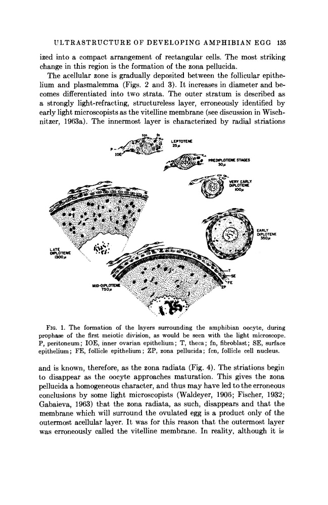
FIG. 1. The formation of the layers surrounding the amphibian oocyte, during
prophase of the first meiotic division, as would be seen with the light microscope.
P, peritoneum; IOE, inner ovarian epithelium; T, theca; fn, fibroblast; SE, surface
epithelium; FE, follicle epithelium; ZP, zona pellucida; fen, follicle cell nucleus.
and is known, therefore, as the zona radiata (Fig. 4). The striations begin
to disappear as the oocyte approaches maturation. This gives the zona
pellucida a homogeneous character, and thus may have led to the erroneous
conclusions by some light microscopists (Waldeyer, 1906; Fischer, 1932;
Gabaieva, 1963) that the zona radiata, as such, disappears and that the
membrane which will surround the ovulated egg is a product only of the
outermost acellular layer. It was for this reason that the outermost layer
was erroneously called the vitelline membrane. In reality, although it is
ized into a compact arrangement of rectangular cells. The most striking
change in this region is the formation of the zona pellucida.
The acellular zone is gradually deposited between the follicular epithelium and plasmalemma (Figs. 2 and 3). It increases in diameter and becomes differentiated into two strata. The outer stratum is described as
a strongly light-refracting, structureless layer, erroneously identified by
early light microscopists as the vitelline membrane (see discussion in Wischnitzer, 1963a). The innermost layer is characterized by radial striatums
FIG. 1. The formation of the layers surrounding the amphibian oocyte, during
prophase of the first meiotic division, as would be seen with the light microscope.
P, peritoneum; IOE, inner ovarian epithelium; T, theca; fn, fibroblast; SE, surface
epithelium; FE, follicle epithelium; ZP, zona pellucida; fen, follicle cell nucleus.
and is known, therefore, as the zona radiata (Fig. 4). The striations begin
to disappear as the oocyte approaches maturation. This gives the zona
pellucida a homogeneous character, and thus may have led to the erroneous
conclusions by some light microscopists (Waldeyer, 1906; Fischer, 1932;
Gabaieva, 1963) that the zona radiata, as such, disappears and that the
membrane which will surround the ovulated egg is a product only of the
outermost acellular layer. It was for this reason that the outermost layer
was erroneously called the vitelline membrane. In reality, although it is