

TRANSPLANTATION OF CELL NUCLEI
25
B. Reversible Changes in Living Nuclei
A functional demonstration of reversible gene activity is provided by
the normal development of transplant-embryos following the transfer
of nuclei from differentiated cells as was described in the last section.
This is now supported by the following cytological and chemical
evidence.
1. Transfer of Amphibian Nuclei to Egg Cytoplasm
The cytological consequences of nuclear transplantation in Xenopus
have been illustrated previously (Gurdon, 1960a). In Rana pipiens
Subtelny and Bradt (1963) have reported a two- to threefold increase
in nuclear volume 30 min after transplantation. In Xenopus the swelling
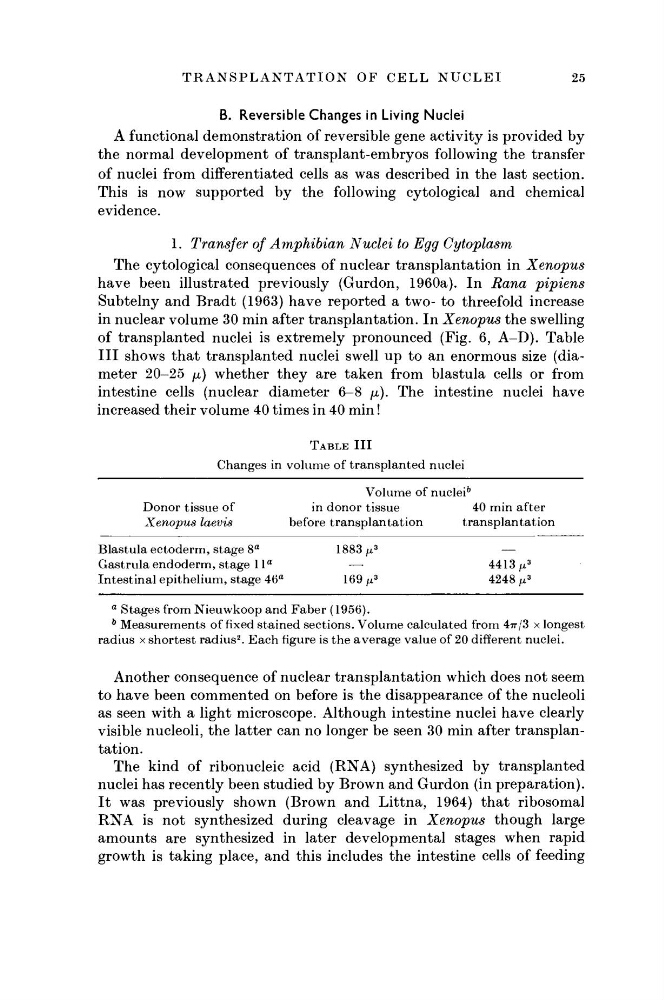
of transplanted nuclei is extremely pronounced (Fig. 6, A-D). Table
III shows that transplanted nuclei swell up to an enormous size (diameter 20-25 μ) whether they are taken from blastula cells or from
intestine cells (nuclear diameter 6-8 μ). The intestine nuclei have
increased their volume 40 times in 40 min !
TABLE III
Changes in volume of transplanted nuclei
Volume of nuclei
5
Donor tissue of
in donor tissue
40 min after
Xenopus laevis
before transplantation
transplantation
Blastula ectoderm, stage 8
a
1883 μ
3
—
Gastrula endoderm, stage 1 l
a
—
4413/x
3
Intestinal epithelium, stage 46
a
169/x
3
4248 μ
3
a Stages from Nieuwkoop and Faber (1956).
b Measurements of fixed stained sections. Volume calculated from 47r/3 X longest
radius x shortest radius
2 . Each figure is the average value of 20 different nuclei.
Another consequence of nuclear transplantation which does not seem
to have been commented on before is the disappearance of the nucleoli
as seen with a light microscope. Although intestine nuclei have clearly
visible nucleoli, the latter can no longer be seen 30 min after transplantation.
The kind of ribonucleic acid (RNA) synthesized by transplanted
nuclei has recently been studied by Brown and Gurdon (in preparation).
It was previously shown (Brown and Littna, 1964) that ribosomal
RNA is not synthesized during cleavage in Xenopus though large
amounts are synthesized in later developmental stages when rapid
growth is taking place, and this includes the intestine cells of feeding
25
B. Reversible Changes in Living Nuclei
A functional demonstration of reversible gene activity is provided by
the normal development of transplant-embryos following the transfer
of nuclei from differentiated cells as was described in the last section.
This is now supported by the following cytological and chemical
evidence.
1. Transfer of Amphibian Nuclei to Egg Cytoplasm
The cytological consequences of nuclear transplantation in Xenopus
have been illustrated previously (Gurdon, 1960a). In Rana pipiens
Subtelny and Bradt (1963) have reported a two- to threefold increase
in nuclear volume 30 min after transplantation. In Xenopus the swelling
of transplanted nuclei is extremely pronounced (Fig. 6, A-D). Table
III shows that transplanted nuclei swell up to an enormous size (diameter 20-25 μ) whether they are taken from blastula cells or from
intestine cells (nuclear diameter 6-8 μ). The intestine nuclei have
increased their volume 40 times in 40 min !
TABLE III
Changes in volume of transplanted nuclei
Volume of nuclei
5
Donor tissue of
in donor tissue
40 min after
Xenopus laevis
before transplantation
transplantation
Blastula ectoderm, stage 8
a
1883 μ
3
—
Gastrula endoderm, stage 1 l
a
—
4413/x
3
Intestinal epithelium, stage 46
a
169/x
3
4248 μ
3
a Stages from Nieuwkoop and Faber (1956).
b Measurements of fixed stained sections. Volume calculated from 47r/3 X longest
radius x shortest radius
2 . Each figure is the average value of 20 different nuclei.
Another consequence of nuclear transplantation which does not seem
to have been commented on before is the disappearance of the nucleoli
as seen with a light microscope. Although intestine nuclei have clearly
visible nucleoli, the latter can no longer be seen 30 min after transplantation.
The kind of ribonucleic acid (RNA) synthesized by transplanted
nuclei has recently been studied by Brown and Gurdon (in preparation).
It was previously shown (Brown and Littna, 1964) that ribosomal
RNA is not synthesized during cleavage in Xenopus though large
amounts are synthesized in later developmental stages when rapid
growth is taking place, and this includes the intestine cells of feeding