

Y O L K OF T H E H E N ' S EGG
255
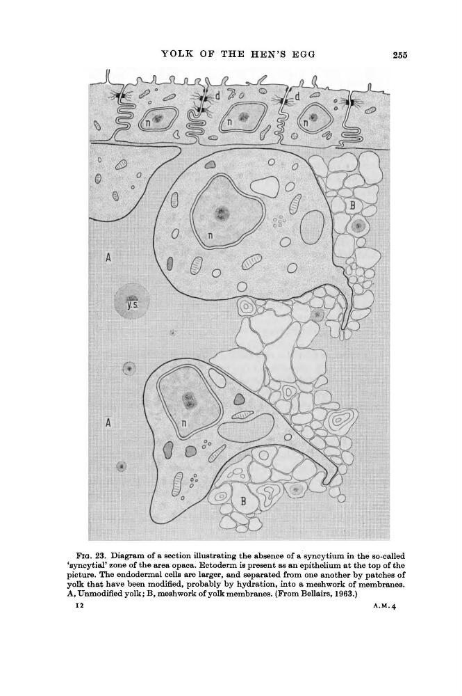
FIG. 23. Diagram of a section illustrating the absence of a syncytium in the so-called
'syncytial' zone of the area opaca. Ectoderm is present as an epithelium at the top of the
picture. The endodermal cells are larger, and separated from one another by patches of
yolk that have been modified, probably by hydration, into a meshwork of membranes.
A, Unmodified yolk; B, meshwork of yolk membranes. (From Bellairs, 1963.)
12
A.M.4
255
FIG. 23. Diagram of a section illustrating the absence of a syncytium in the so-called
'syncytial' zone of the area opaca. Ectoderm is present as an epithelium at the top of the
picture. The endodermal cells are larger, and separated from one another by patches of
yolk that have been modified, probably by hydration, into a meshwork of membranes.
A, Unmodified yolk; B, meshwork of yolk membranes. (From Bellairs, 1963.)
12
A.M.4