

246
RUTH BELLAIRS
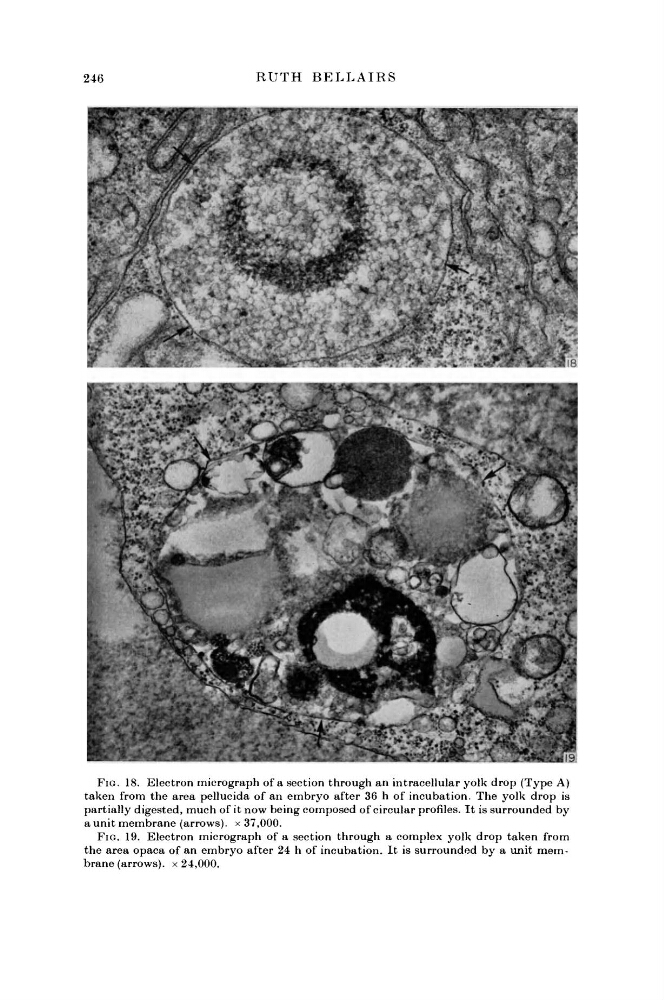
FIG. 18. Electron micrograph of a section through an intracellular yolk drop (Type A)
taken from the area pellucida of an embryo after 36 h of incubation. The yolk drop is
partially digested, much of it now being composed of circular profiles. It is surrounded by
a unit membrane (arrows), x 37,000.
FIG. 19. Electron micrograph of a section through a complex yolk drop taken from
the area opaca of an embryo after 24 h of incubation. It is surrounded by a unit membrane (arrows), x 24,000.
RUTH BELLAIRS
FIG. 18. Electron micrograph of a section through an intracellular yolk drop (Type A)
taken from the area pellucida of an embryo after 36 h of incubation. The yolk drop is
partially digested, much of it now being composed of circular profiles. It is surrounded by
a unit membrane (arrows), x 37,000.
FIG. 19. Electron micrograph of a section through a complex yolk drop taken from
the area opaca of an embryo after 24 h of incubation. It is surrounded by a unit membrane (arrows), x 24,000.