

Y O L K OF T H E H E N ' S E G G
243
During its final growth phase the oocyte ceases to be spherical and
becomes longer in one direction. The follicle becomes so distended that
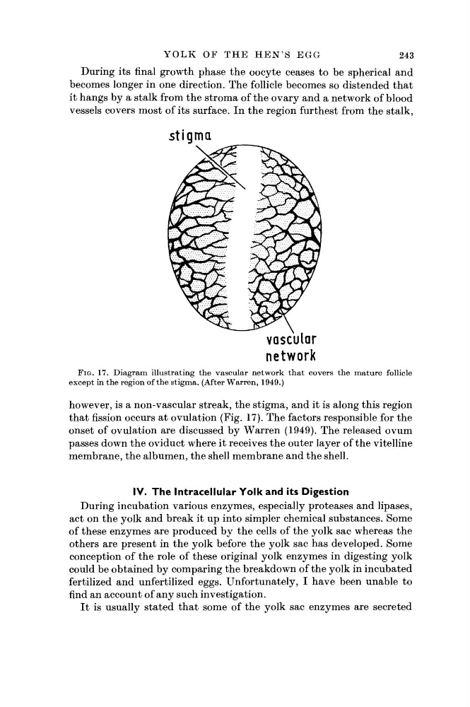
it hangs by a stalk from the stroma of the ovary and a network of blood
vessels covers most of its surface. In the region furthest from the stalk,
stigma
FIG. 17. Diagram illustrating the vascular network that covers the mature follicle
except in the region of the stigma. (After Warren, 1949.)
however, is a non-vascular streak, the stigma, and it is along this region
that fission occurs at ovulation (Fig. 17). The factors responsible for the
onset of ovulation are discussed by Warren (1949). The released ovum
passes down the oviduct where it receives the outer layer of the vitelline
membrane, the albumen, the shell membrane and the shell.
IV. The Intracellular Yolk and its Digestion
During incubation various enzymes, especially proteases and Upases,
act on the yolk and break it up into simpler chemical substances. Some
of these enzymes are produced by the cells of the yolk sac whereas the
others are present in the yolk before the yolk sac has developed. Some
conception of the role of these original yolk enzymes in digesting yolk
could be obtained by comparing the breakdown of the yolk in incubated
fertilized and unfertilized eggs. Unfortunately, I have been unable to
find an account of any such investigation.
It is usually stated that some of the yolk sac enzymes are secreted
243
During its final growth phase the oocyte ceases to be spherical and
becomes longer in one direction. The follicle becomes so distended that
it hangs by a stalk from the stroma of the ovary and a network of blood
vessels covers most of its surface. In the region furthest from the stalk,
stigma
FIG. 17. Diagram illustrating the vascular network that covers the mature follicle
except in the region of the stigma. (After Warren, 1949.)
however, is a non-vascular streak, the stigma, and it is along this region
that fission occurs at ovulation (Fig. 17). The factors responsible for the
onset of ovulation are discussed by Warren (1949). The released ovum
passes down the oviduct where it receives the outer layer of the vitelline
membrane, the albumen, the shell membrane and the shell.
IV. The Intracellular Yolk and its Digestion
During incubation various enzymes, especially proteases and Upases,
act on the yolk and break it up into simpler chemical substances. Some
of these enzymes are produced by the cells of the yolk sac whereas the
others are present in the yolk before the yolk sac has developed. Some
conception of the role of these original yolk enzymes in digesting yolk
could be obtained by comparing the breakdown of the yolk in incubated
fertilized and unfertilized eggs. Unfortunately, I have been unable to
find an account of any such investigation.
It is usually stated that some of the yolk sac enzymes are secreted