

232
R U T H B E L L A I R S
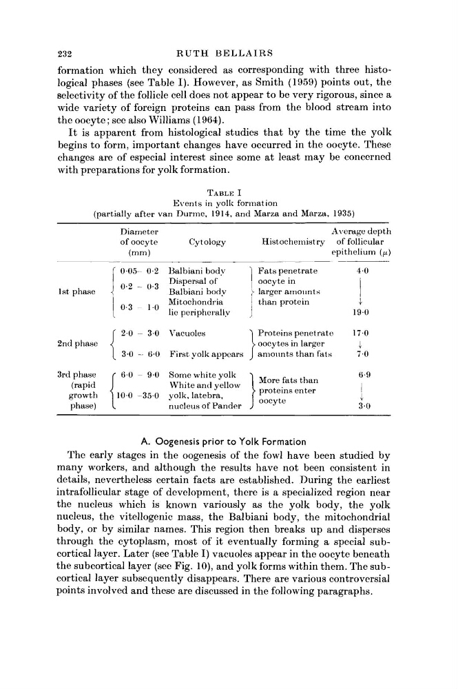
formation which they considered as corresponding with three histological phases (see Table I). However, as Smith (1959) points out, the
selectivity of the follicle cell does not appear to be very rigorous, since a
wide variety of foreign proteins can pass from the blood stream into
the oocyte; see also Williams (1964).
It is apparent from histological studies that by the time the yolk
begins to form, important changes have occurred in the oocyte. These
changes are of especial interest since some at least may be concerned
with preparations for yolk formation.
T A B L E I
E v e n t s in yolk formation
(partially after van D u r m e , 1914, and Marza a n d Marza, 1935)
Diameter
Average d e p t h
of oocyte
Cytology
Histochemistry
of follicular
(mm)
epithelium (μ)
1st phase
2nd phase
3rd phase
(rapid
growth
phase)
0 0 5 - 0-2 B a l b i a n i b o d y
Dispersal of
Balbiani body
Mitochondria
lie peripherally
0-2
0-3
0-3
1 0
Ί F a t s penetrate
oocyte in
Ϊ larger a m o u n t s
t h a n protein
2 0 - 3-0 Vacuoles
3-0 - 6 0 First yolk appears
6 0 - 9 0 Some white yolk
W h i t e and yellow
1 0 0 - 3 5 0 yolk, latebra,
nucleus of P a n d e r
Proteins penetrate
> oocytes in larger
a m o u n t s t h a n fats
More fats t h a n
proteins enter
oocyte
4 0
1 9 0
170
I
70
6-9
I
I
30
A. Oogenesis prior to Yolk Formation
The early stages in the oogenesis of the fowl have been studied by
many workers, and although the results have not been consistent in
details, nevertheless certain facts are established. During the earliest
intraf ollicular stage of development, there is a specialized region near
the nucleus which is known variously as the yolk body, the yolk
nucleus, the vitellogenic mass, the Balbiani body, the mitochondrial
body, or by similar names. This region then breaks up and disperses
through the cytoplasm, most of it eventually forming a special subcortical layer. Later (see Table I) vacuoles appear in the oocyte beneath
the subcortical layer (see Fig. 10), and yolk forms within them. The subcortical layer subsequently disappears. There are various controversial
points involved and these are discussed in the following paragraphs.
R U T H B E L L A I R S
formation which they considered as corresponding with three histological phases (see Table I). However, as Smith (1959) points out, the
selectivity of the follicle cell does not appear to be very rigorous, since a
wide variety of foreign proteins can pass from the blood stream into
the oocyte; see also Williams (1964).
It is apparent from histological studies that by the time the yolk
begins to form, important changes have occurred in the oocyte. These
changes are of especial interest since some at least may be concerned
with preparations for yolk formation.
T A B L E I
E v e n t s in yolk formation
(partially after van D u r m e , 1914, and Marza a n d Marza, 1935)
Diameter
Average d e p t h
of oocyte
Cytology
Histochemistry
of follicular
(mm)
epithelium (μ)
1st phase
2nd phase
3rd phase
(rapid
growth
phase)
0 0 5 - 0-2 B a l b i a n i b o d y
Dispersal of
Balbiani body
Mitochondria
lie peripherally
0-2
0-3
0-3
1 0
Ί F a t s penetrate
oocyte in
Ϊ larger a m o u n t s
t h a n protein
2 0 - 3-0 Vacuoles
3-0 - 6 0 First yolk appears
6 0 - 9 0 Some white yolk
W h i t e and yellow
1 0 0 - 3 5 0 yolk, latebra,
nucleus of P a n d e r
Proteins penetrate
> oocytes in larger
a m o u n t s t h a n fats
More fats t h a n
proteins enter
oocyte
4 0
1 9 0
170
I
70
6-9
I
I
30
A. Oogenesis prior to Yolk Formation
The early stages in the oogenesis of the fowl have been studied by
many workers, and although the results have not been consistent in
details, nevertheless certain facts are established. During the earliest
intraf ollicular stage of development, there is a specialized region near
the nucleus which is known variously as the yolk body, the yolk
nucleus, the vitellogenic mass, the Balbiani body, the mitochondrial
body, or by similar names. This region then breaks up and disperses
through the cytoplasm, most of it eventually forming a special subcortical layer. Later (see Table I) vacuoles appear in the oocyte beneath
the subcortical layer (see Fig. 10), and yolk forms within them. The subcortical layer subsequently disappears. There are various controversial
points involved and these are discussed in the following paragraphs.