

YOLK OF THE HEN'S EGG
229
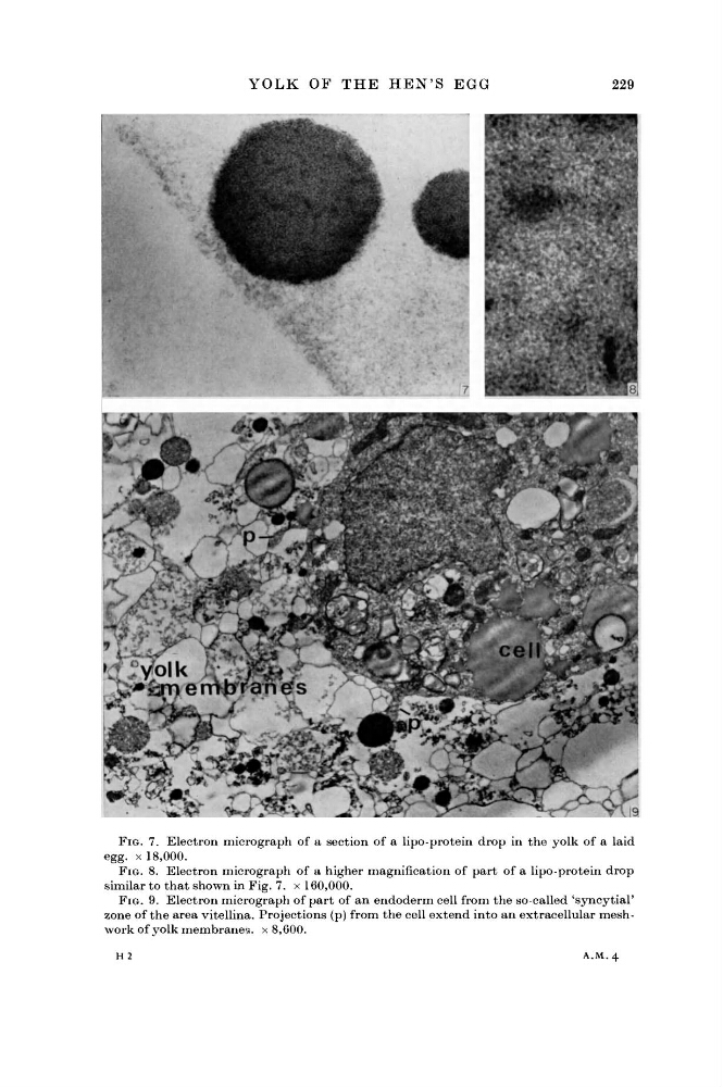
FIG. 7. Electron micrograph of a section of a lipo-protein drop in the yolk of a laid
egg. x 18,000.
FIG. 8. Electron micrograph of a higher magnification of part of a lipo-protein drop
similar to that shown in Fig. 7. x 160,000.
FIG. 9. Electron micrograph of part of an endoderm cell from the so-called 'syncytial'
zone of the area vitellina. Projections (p) from the cell extend into an extracellular meshwork of yolk membranes, x 8,600.
H 2
A . M . 4
229
FIG. 7. Electron micrograph of a section of a lipo-protein drop in the yolk of a laid
egg. x 18,000.
FIG. 8. Electron micrograph of a higher magnification of part of a lipo-protein drop
similar to that shown in Fig. 7. x 160,000.
FIG. 9. Electron micrograph of part of an endoderm cell from the so-called 'syncytial'
zone of the area vitellina. Projections (p) from the cell extend into an extracellular meshwork of yolk membranes, x 8,600.
H 2
A . M . 4