

YOLK OF THE HEN'S EGG
221
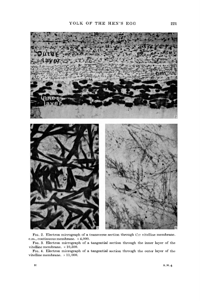
FIG. 2. Electron micrograph of a transverse section through the vitelline membrane.
c m . , continuous membrane, x 4,000.
FIG. 3. Electron micrograph of a tangential section through the inner layer of the
vitelline membrane, x 10,500.
FIG. 4. Electron micrograph of a tangential section through the outer layer of the
vitelline membrane, x 11,000.
H
A.M.4
221
FIG. 2. Electron micrograph of a transverse section through the vitelline membrane.
c m . , continuous membrane, x 4,000.
FIG. 3. Electron micrograph of a tangential section through the inner layer of the
vitelline membrane, x 10,500.
FIG. 4. Electron micrograph of a tangential section through the outer layer of the
vitelline membrane, x 11,000.
H
A.M.4