

YOLK OF THE HEN'S EGG
219
nucleus of
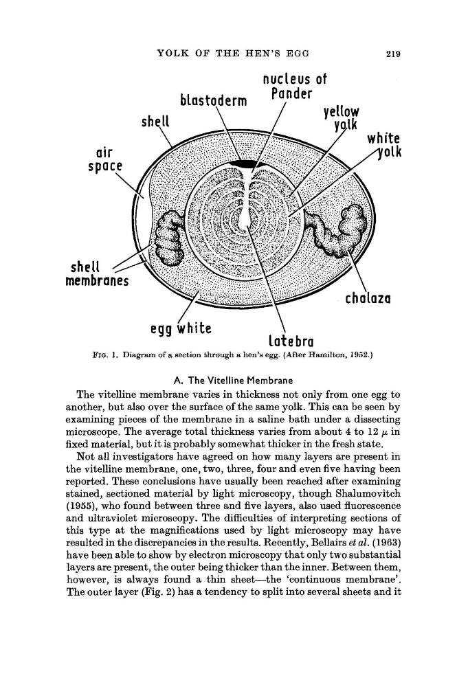
FIG. 1. Diagram of a section through a hen's egg. (After Hamilton, 1952.)
A. The Vitelline Membrane
The vitelline membrane varies in thickness not only from one egg to
another, but also over the surface of the same yolk. This can be seen by
examining pieces of the membrane in a saline bath under a dissecting
microscope. The average total thickness varies from about 4 to 12 μ in
fixed material, but it is probably somewhat thicker in the fresh state.
Not all investigators have agreed on how many layers are present in
the vitelline membrane, one, two, three, four and even five having been
reported. These conclusions have usually been reached after examining
stained, sectioned material by light microscopy, though Shalumovitch
(1955), who found between three and five layers, also used fluorescence
and ultraviolet microscopy. The difficulties of interpreting sections of
this type at the magnifications used by light microscopy may have
resulted in the discrepancies in the results. Recently, Bellairs et al. (1963)
have been able to show by electron microscopy that only two substantial
layers are present, the outer being thicker than the inner. Between them,
however, is always found a thin sheet—the 'continuous membrane'.
The outer layer (Fig. 2) has a tendency to split into several sheets and it
219
nucleus of
FIG. 1. Diagram of a section through a hen's egg. (After Hamilton, 1952.)
A. The Vitelline Membrane
The vitelline membrane varies in thickness not only from one egg to
another, but also over the surface of the same yolk. This can be seen by
examining pieces of the membrane in a saline bath under a dissecting
microscope. The average total thickness varies from about 4 to 12 μ in
fixed material, but it is probably somewhat thicker in the fresh state.
Not all investigators have agreed on how many layers are present in
the vitelline membrane, one, two, three, four and even five having been
reported. These conclusions have usually been reached after examining
stained, sectioned material by light microscopy, though Shalumovitch
(1955), who found between three and five layers, also used fluorescence
and ultraviolet microscopy. The difficulties of interpreting sections of
this type at the magnifications used by light microscopy may have
resulted in the discrepancies in the results. Recently, Bellairs et al. (1963)
have been able to show by electron microscopy that only two substantial
layers are present, the outer being thicker than the inner. Between them,
however, is always found a thin sheet—the 'continuous membrane'.
The outer layer (Fig. 2) has a tendency to split into several sheets and it