

180
H E N R I E T T E HERLANT-MEEWIS
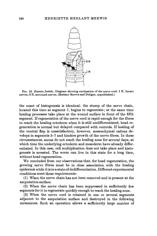
FIG. 19. Eieenia foetida. Diagram showing extirpation of the nerve cord. I N, Intact
nerves; S N, sectioned nerves. (Herlant-Meewis and Deligne, unpublished.)
the onset of histogenesis is identical: the stump of the nerve chain,
located this time at segment 7, begins to regenerate; at the same time
healing processes take place at the wound surface in front of the fifth
segment. If regeneration of the nerve cord is rapid enough for the fibres
to reach the healing ectoderm when it is still undifferentiated, head regeneration is normal but delayed compared with controls. If healing of
the ventral flap is unsatisfactory, however, mesenchymal callous develops in segments 5-7 and hinders growth of the nerve fibres. In these
circumstances, axons do not reach the healing zone for several days, at
which time the underlying ectoderm and mesoderm have already differentiated. In this case, cell multiplication does not take place and histogenesis is arrested. The worm can live in this state for a long time,
without head regeneration.
We concluded from our observations that, for head regeneration, the
growing nerve fibres must be in close association with the healing
epidermis while it is in a state of dedifferentiation. Different experimental
conditions meet these requirements :
(1) When the nerve chain has not been removed and is present at the
amputation surface.
(2) When the nerve chain has been suppressed in sufficiently few
segments for it to regenerate quickly enough to reach the healing zone.
(3) When the nerve cord is retained in one or several segments
adjacent to the amputation surface and destroyed in the following
metameres. Such an operation allows a sufficiently large number of
H E N R I E T T E HERLANT-MEEWIS
FIG. 19. Eieenia foetida. Diagram showing extirpation of the nerve cord. I N, Intact
nerves; S N, sectioned nerves. (Herlant-Meewis and Deligne, unpublished.)
the onset of histogenesis is identical: the stump of the nerve chain,
located this time at segment 7, begins to regenerate; at the same time
healing processes take place at the wound surface in front of the fifth
segment. If regeneration of the nerve cord is rapid enough for the fibres
to reach the healing ectoderm when it is still undifferentiated, head regeneration is normal but delayed compared with controls. If healing of
the ventral flap is unsatisfactory, however, mesenchymal callous develops in segments 5-7 and hinders growth of the nerve fibres. In these
circumstances, axons do not reach the healing zone for several days, at
which time the underlying ectoderm and mesoderm have already differentiated. In this case, cell multiplication does not take place and histogenesis is arrested. The worm can live in this state for a long time,
without head regeneration.
We concluded from our observations that, for head regeneration, the
growing nerve fibres must be in close association with the healing
epidermis while it is in a state of dedifferentiation. Different experimental
conditions meet these requirements :
(1) When the nerve chain has not been removed and is present at the
amputation surface.
(2) When the nerve chain has been suppressed in sufficiently few
segments for it to regenerate quickly enough to reach the healing zone.
(3) When the nerve cord is retained in one or several segments
adjacent to the amputation surface and destroyed in the following
metameres. Such an operation allows a sufficiently large number of