

TRANSPLANTATION OF CELL NUCLEI
15
happens to be in an unfavourable stage of mitosis at the time of transplantation. From what we know at present these different kinds of
abnormal or deficient cleavage are caused by technical factors or the
mitotic state of the donor nucleus, and this would account for their
increased frequency following the injection of nuclei from tadpole cells
compared to blastula nuclei. For these reasons we can regard the
development of only those recipient eggs which cleave regularly as
showing the developmental capacity of transplanted nuclei.
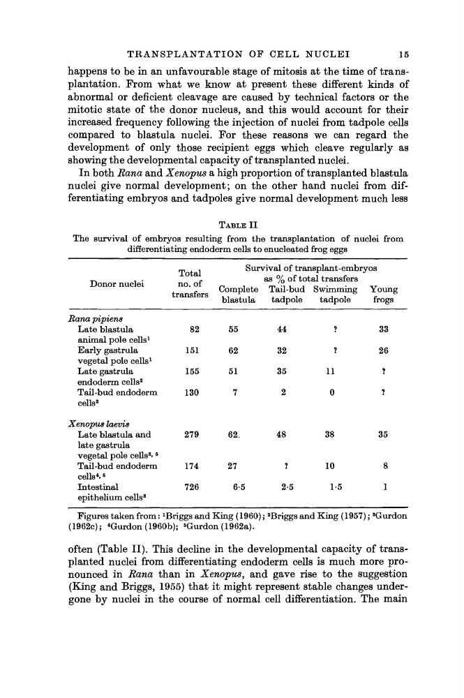
In both Rana and Xenopus a high proportion of transplanted blastula
nuclei give normal development; on the other hand nuclei from differentiating embryos and tadpoles give normal development much less
TABLE I I
The survival of embryos resulting from the transplantation of nuclei from
differentiating endoderm cells to enucleated frog eggs
Donor nuclei
Ranapipiens
Late blastula
animal pole cells
1
Early gastrula
vegetal pole cells
1
Late gastrula
endoderm cells
2
Tail-bud endoderm
cells
2
Xenopus laevis
Late blastula and
late gastrula
vegetal pole cells
3 »
5
Tail-bud endoderm
cells
4 »
5
Intestinal
epithelium cells
3
Total
no. of
ül cUlolUl o
82
151
155
130
279
174
726
Survival of transplant-embryos
Complete
blastula
55
62
51
7
62.
27
6-5
as % of total transfers
Tail-bud Swimming
tadpole
44
32
35
2
48
?
2-5
tadpole
?
?
11
0
38
10
1-5
Young
frogs
33
26
?
?
35
8
1
Figures taken from :
1 Briggs and King (1960) ;
2 Briggs and King (1957) ;
8 Gurdon
(1962c);
4 Gurdon (1960b);
5 Gurdon (1962a).
often (Table II). This decline in the developmental capacity of transplanted nuclei from differentiating endoderm cells is much more pronounced in Rana than in Xenopus, and gave rise to the suggestion
(King and Briggs, 1955) that it might represent stable changes undergone by nuclei in the course of normal cell differentiation. The main
15
happens to be in an unfavourable stage of mitosis at the time of transplantation. From what we know at present these different kinds of
abnormal or deficient cleavage are caused by technical factors or the
mitotic state of the donor nucleus, and this would account for their
increased frequency following the injection of nuclei from tadpole cells
compared to blastula nuclei. For these reasons we can regard the
development of only those recipient eggs which cleave regularly as
showing the developmental capacity of transplanted nuclei.
In both Rana and Xenopus a high proportion of transplanted blastula
nuclei give normal development; on the other hand nuclei from differentiating embryos and tadpoles give normal development much less
TABLE I I
The survival of embryos resulting from the transplantation of nuclei from
differentiating endoderm cells to enucleated frog eggs
Donor nuclei
Ranapipiens
Late blastula
animal pole cells
1
Early gastrula
vegetal pole cells
1
Late gastrula
endoderm cells
2
Tail-bud endoderm
cells
2
Xenopus laevis
Late blastula and
late gastrula
vegetal pole cells
3 »
5
Tail-bud endoderm
cells
4 »
5
Intestinal
epithelium cells
3
Total
no. of
ül cUlolUl o
82
151
155
130
279
174
726
Survival of transplant-embryos
Complete
blastula
55
62
51
7
62.
27
6-5
as % of total transfers
Tail-bud Swimming
tadpole
44
32
35
2
48
?
2-5
tadpole
?
?
11
0
38
10
1-5
Young
frogs
33
26
?
?
35
8
1
Figures taken from :
1 Briggs and King (1960) ;
2 Briggs and King (1957) ;
8 Gurdon
(1962c);
4 Gurdon (1960b);
5 Gurdon (1962a).
often (Table II). This decline in the developmental capacity of transplanted nuclei from differentiating endoderm cells is much more pronounced in Rana than in Xenopus, and gave rise to the suggestion
(King and Briggs, 1955) that it might represent stable changes undergone by nuclei in the course of normal cell differentiation. The main