

R E G E N E R A T I O N IN ANNELIDS
163
tion. Stephan-Dubois (1956) counted neoblasts during cephalic regeneration in Lumbriculus variegatus. Activation of septal cells began
within an hour of amputation ; 24 h later the neoblasts had migrated
and had gathered in the segments adjacent to the wound. Cell division
began on the 3rd day.
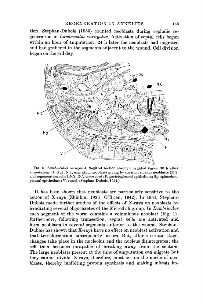
FIG. 8. Lumbriculus variegatus. Sagittal section through pygidial region 30 h after
amputation. G, Gut; N 1, migrating neoblasts giving by division smaller neoblasts (N 2)
and regeneration cells (RC) ; NC, nerve cord; P, parietopleural epithelium; Sp, splanchnopleural epithelium; V, vessel. (Stephan-Dubois, 1954.)
I t has been shown that neoblasts are particularly sensitive to the
action of X-rays (Zhinkin, 1936; O'Brien, 1942). In 1954, StephanDubois made further studies of the effects of X-rays on neoblasts by
irradiating several oligochaetes of the Microdrili group. In Lumbriculus
each segment of the worm contains a voluminous neoblast (Fig. 1);
furthermore, following transection, septal cells are activated and
form neoblasts in several segments anterior to the wound. StephanDubois has shown that X-rays have no effect on neoblast activation and
that transformation subsequently occurs. But, after a certain stage,
changes take place in the nucleolus and the nucleus disintegrates; the
cell then becomes incapable of breaking away from the septum.
The large neoblasts present at the time of amputation can migrate but
they cannot divide. X-rays, therefore, must act on the nuclei of neoblasts, thereby inhibiting protein synthesis and making mitosis im-
163
tion. Stephan-Dubois (1956) counted neoblasts during cephalic regeneration in Lumbriculus variegatus. Activation of septal cells began
within an hour of amputation ; 24 h later the neoblasts had migrated
and had gathered in the segments adjacent to the wound. Cell division
began on the 3rd day.
FIG. 8. Lumbriculus variegatus. Sagittal section through pygidial region 30 h after
amputation. G, Gut; N 1, migrating neoblasts giving by division smaller neoblasts (N 2)
and regeneration cells (RC) ; NC, nerve cord; P, parietopleural epithelium; Sp, splanchnopleural epithelium; V, vessel. (Stephan-Dubois, 1954.)
I t has been shown that neoblasts are particularly sensitive to the
action of X-rays (Zhinkin, 1936; O'Brien, 1942). In 1954, StephanDubois made further studies of the effects of X-rays on neoblasts by
irradiating several oligochaetes of the Microdrili group. In Lumbriculus
each segment of the worm contains a voluminous neoblast (Fig. 1);
furthermore, following transection, septal cells are activated and
form neoblasts in several segments anterior to the wound. StephanDubois has shown that X-rays have no effect on neoblast activation and
that transformation subsequently occurs. But, after a certain stage,
changes take place in the nucleolus and the nucleus disintegrates; the
cell then becomes incapable of breaking away from the septum.
The large neoblasts present at the time of amputation can migrate but
they cannot divide. X-rays, therefore, must act on the nuclei of neoblasts, thereby inhibiting protein synthesis and making mitosis im-