

42
J. CLAVERT
hours, during which it acquires its shell. During its stay in the uterus,
the egg rotates about its long axis, always in a clockwise direction,
under the influence of the uterine muscle. According to Conrad and
Philipps (1938) this rotation, with a concomitant imbibition of fluid,
may bring about the elaboration of the layers of liquid albumen. The
albumen sac however clings to the shell and the yolk at either end. Thus
the albuminous ligaments and the chalazae are formed.
In the uterus, the egg membranes, connected with each other by the
albuminous ligaments, always rotate in the same direction. Inside, the
yolk remains nearly stationary. The germ, lying close to the albuminous
sac, rubs against it; and the chalazae, which are at first a region where
the albuminous sac clings to the yolk, begin to coil, the spiral becoming
more and more apparent as long as the egg remains in the uterus. The
study of the coiling of the chalazae, as we shall discuss later, may give
fruitful information as to the behaviour of the egg during its stay in the
uterus.
C. Relation between the Position of the Embryo and the
Direction of Coiling of the Chalazae
The chalazae are formed while the egg is in the uterus. When the egg
reaches the isthmus the yolk is not mobile within its membranes; and
when the inner layer of liquid albumen is formed, the yolk still sticks to
the albuminous sac at the poles facing the blunt and sharp ends of the
egg, these adhesions being the chalazae. When the egg membranes
rotate in the uterus, the yolk remains nearly motionless, so that the
chalazae become coiled. Later, this torsion extends to the mucin fibres
of the albuminous sac, and the chalazae coil more closely. The spiral
may be either right-handed (clockwise) or left-handed (anticlockwise).
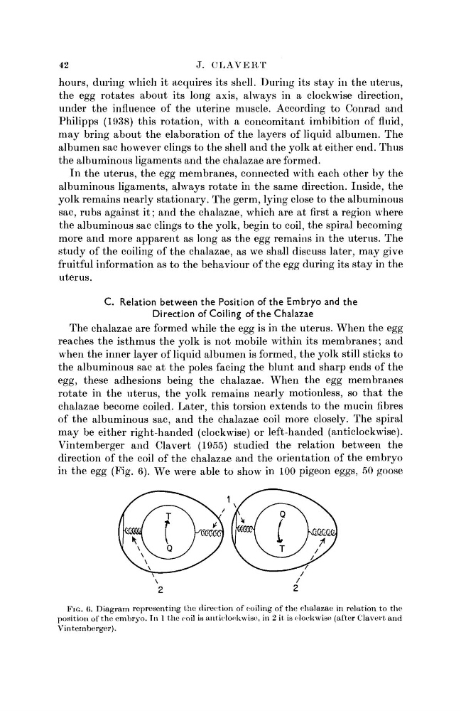
Vintemberger and Clavert (1955) studied the relation between the
direction of the coil of the chalazae and the orientation of the embryo
in the egg (Fig. 6). We were able to show in 100 pigeon eggs, 50 goose
FIG. 6. Diagram representing the direction of coiling of the chalazae in relation to the
position of the embryo. In 1 the coil is anticlockwise, in 2 it is clockwise (after Clavert and
Vintemberger).
J. CLAVERT
hours, during which it acquires its shell. During its stay in the uterus,
the egg rotates about its long axis, always in a clockwise direction,
under the influence of the uterine muscle. According to Conrad and
Philipps (1938) this rotation, with a concomitant imbibition of fluid,
may bring about the elaboration of the layers of liquid albumen. The
albumen sac however clings to the shell and the yolk at either end. Thus
the albuminous ligaments and the chalazae are formed.
In the uterus, the egg membranes, connected with each other by the
albuminous ligaments, always rotate in the same direction. Inside, the
yolk remains nearly stationary. The germ, lying close to the albuminous
sac, rubs against it; and the chalazae, which are at first a region where
the albuminous sac clings to the yolk, begin to coil, the spiral becoming
more and more apparent as long as the egg remains in the uterus. The
study of the coiling of the chalazae, as we shall discuss later, may give
fruitful information as to the behaviour of the egg during its stay in the
uterus.
C. Relation between the Position of the Embryo and the
Direction of Coiling of the Chalazae
The chalazae are formed while the egg is in the uterus. When the egg
reaches the isthmus the yolk is not mobile within its membranes; and
when the inner layer of liquid albumen is formed, the yolk still sticks to
the albuminous sac at the poles facing the blunt and sharp ends of the
egg, these adhesions being the chalazae. When the egg membranes
rotate in the uterus, the yolk remains nearly motionless, so that the
chalazae become coiled. Later, this torsion extends to the mucin fibres
of the albuminous sac, and the chalazae coil more closely. The spiral
may be either right-handed (clockwise) or left-handed (anticlockwise).
Vintemberger and Clavert (1955) studied the relation between the
direction of the coil of the chalazae and the orientation of the embryo
in the egg (Fig. 6). We were able to show in 100 pigeon eggs, 50 goose
FIG. 6. Diagram representing the direction of coiling of the chalazae in relation to the
position of the embryo. In 1 the coil is anticlockwise, in 2 it is clockwise (after Clavert and
Vintemberger).