

40
J. CLAVERT
and Romanoff (1949), Scott and Huang (1941), Warren and Scott
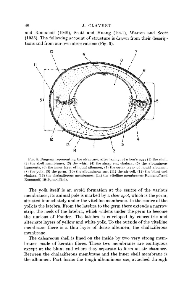
(1935). The following account of structure is drawn from their descriptions and from our own observations (Fig. 5).
FIG. 5. Diagram representing the structure, after laying, of a hen's egg; (1) the shell,
(2) the shell membranes, (3) the whirl, (4) the sharp end chalaza, (5) the albuminous
ligaments, (6) the inner layer of liquid albumen, (7) the outer layer of liquid albumen,
(8) the yolk, (9) the germ, (10) the albuminous sac, (11) the air cell, (12) the blunt end
chalaza, (13) the chalaziferous membranes, (14) the vitelline membranes (Romanoff and
Romanoff, 1949, modified).
The yolk itself is an ovoid formation at the centre of the various
membranes; its animal pole is marked by a clear spot, which is the germ,
situated immediately under the vitelline membrane. In the centre of the
yolk is the latebra. From the latebra to the germ there extends a narrow
strip, the neck of the latebra, which widens under the germ to become
the nucleus of Pander. The latebra is enveloped by concentric and
alternate layers of yellow and white yolk. To the outside of the vitelline
membrane there is a thin layer of dense albumen, the chalaziferous
membrane.
The calcareous shell is lined on the inside by two very strong membranes made of keratin fibres. These two membranes are contiguous
except at the blunt end where they separate to form an air chamber.
Between the chalaziferous membrane and the inner shell membrane is
the albumen. Part forms the tough albuminous sac, attached through
J. CLAVERT
and Romanoff (1949), Scott and Huang (1941), Warren and Scott
(1935). The following account of structure is drawn from their descriptions and from our own observations (Fig. 5).
FIG. 5. Diagram representing the structure, after laying, of a hen's egg; (1) the shell,
(2) the shell membranes, (3) the whirl, (4) the sharp end chalaza, (5) the albuminous
ligaments, (6) the inner layer of liquid albumen, (7) the outer layer of liquid albumen,
(8) the yolk, (9) the germ, (10) the albuminous sac, (11) the air cell, (12) the blunt end
chalaza, (13) the chalaziferous membranes, (14) the vitelline membranes (Romanoff and
Romanoff, 1949, modified).
The yolk itself is an ovoid formation at the centre of the various
membranes; its animal pole is marked by a clear spot, which is the germ,
situated immediately under the vitelline membrane. In the centre of the
yolk is the latebra. From the latebra to the germ there extends a narrow
strip, the neck of the latebra, which widens under the germ to become
the nucleus of Pander. The latebra is enveloped by concentric and
alternate layers of yellow and white yolk. To the outside of the vitelline
membrane there is a thin layer of dense albumen, the chalaziferous
membrane.
The calcareous shell is lined on the inside by two very strong membranes made of keratin fibres. These two membranes are contiguous
except at the blunt end where they separate to form an air chamber.
Between the chalaziferous membrane and the inner shell membrane is
the albumen. Part forms the tough albuminous sac, attached through