

E X P E R I M E N T S ON EMBRYONIC AORTIC ARCHES 361
after local irradiation of the heart by means of X-rays in early chick
embryos. The meaning of the persistence of the bulboventricular groove
for the development of the transposition of aorta into the right ventricle
has been discussed theoretically by de la Cruz and da Rocha (1956).
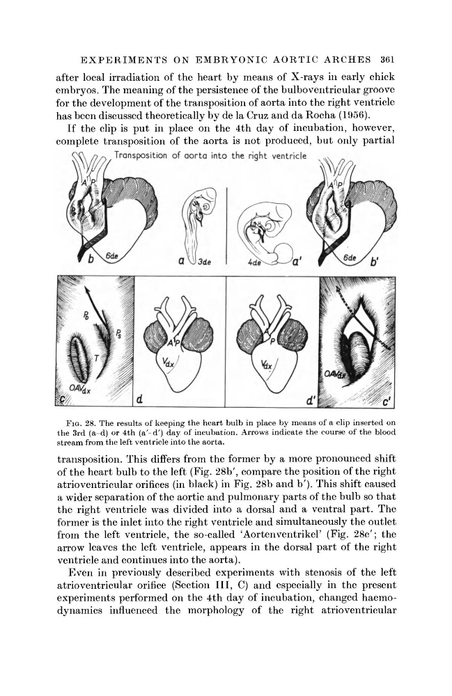
If the clip is put in place on the 4th day of incubation, however,
complete transposition of the aorta is not produced, but only partial
Transposition of aorta into the right ventricle
FIG. 28. The results of keeping the heart bulb in place by means of a clip inserted on
the 3rd (a-d) or 4th (a'- d') day of incubation. Arrows indicate the course of the blood
stream from the left ventricle into the aorta.
transposition. This differs from the former by a more pronounced shift
of the heart bulb to the left (Fig. 28b', compare the position of the right
atrioventricular orifices (in black) in Fig. 28b and b'). This shift caused
a wider separation of the aortic and pulmonary parts of the bulb so that
the right ventricle was divided into a dorsal and a ventral part. The
former is the inlet into the right ventricle and simultaneously the outlet
from the left ventricle, the so-called 'Aortenventrikel' (Fig. 28c'; the
arrow leaves the left ventricle, appears in the dorsal part of the right
ventricle and continues into the aorta).
Even in previously described experiments with stenosis of the left
atrioventricular orifice (Section III, C) and especially in the present
experiments performed on the 4th day of incubation, changed haemodynamics influenced the morphology of the right atrioventricular
after local irradiation of the heart by means of X-rays in early chick
embryos. The meaning of the persistence of the bulboventricular groove
for the development of the transposition of aorta into the right ventricle
has been discussed theoretically by de la Cruz and da Rocha (1956).
If the clip is put in place on the 4th day of incubation, however,
complete transposition of the aorta is not produced, but only partial
Transposition of aorta into the right ventricle
FIG. 28. The results of keeping the heart bulb in place by means of a clip inserted on
the 3rd (a-d) or 4th (a'- d') day of incubation. Arrows indicate the course of the blood
stream from the left ventricle into the aorta.
transposition. This differs from the former by a more pronounced shift
of the heart bulb to the left (Fig. 28b', compare the position of the right
atrioventricular orifices (in black) in Fig. 28b and b'). This shift caused
a wider separation of the aortic and pulmonary parts of the bulb so that
the right ventricle was divided into a dorsal and a ventral part. The
former is the inlet into the right ventricle and simultaneously the outlet
from the left ventricle, the so-called 'Aortenventrikel' (Fig. 28c'; the
arrow leaves the left ventricle, appears in the dorsal part of the right
ventricle and continues into the aorta).
Even in previously described experiments with stenosis of the left
atrioventricular orifice (Section III, C) and especially in the present
experiments performed on the 4th day of incubation, changed haemodynamics influenced the morphology of the right atrioventricular