

314
TH. LENDER
tigrina), implanted in the postpharyngeal region, caused the differentiation of one or two supplementary pharynges in the postpharyngeal
region where the tissues of the host become reorganized by the graft.
They acquired pharyngeal competence. The head graft, then, played the
role of an inducer. Santos assumed that the induction was carried out by
the brain of the graft. In 1936, Chevtchenko confirmed these results on
Planaria lugubris.
Sugino (1940, 1941) studied the induction of the pharynx at different
levels of the body in Planaria (Dugesia) gonocephala. He grafted a head
in the postpharyngeal, pharyngeal, or prepharyngeal region. The first
two regions built up a supplementary pharynx under the influence of the
graft, but the prepharyngeal region was not reorganized and did not
differentiate into pharynx.
Okada and Sugino (1934, 1937) and Okada and Kido (1943) showed
that the prepharyngeal region of Planaria (Dugesia) gonocephala, when
grafted to the tail, also induced differentiation of a pharynx in the
tissues of the host. These experiments conflict with the results of Santos.
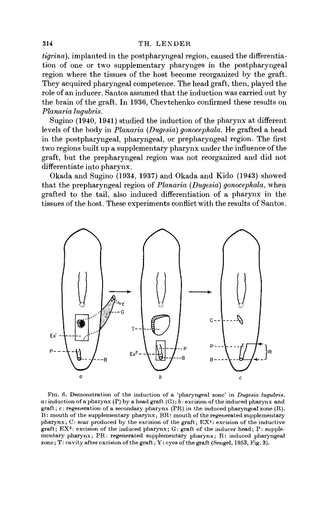
FIG. 6. Demonstration of the induction of a 'pharyngeal zone' in Dugesia lugubris.
a: induction of a pharynx (P) by a head graft (G); b: excision of the induced pharynx and
graft; c: regeneration of a secondary pharynx (PR) in the induced pharyngeal zone (R).
B : mouth of the supplementary pharynx; BR: mouth of the regenerated supplementary
pharynx; C: scar produced by the excision of the graft; E X
1 : excision of the inductive
graft; E X
2 : excision of the induced pharynx; G: graft of the inducer head; P : supplementary pharynx; P R : regenerated supplementary pharynx; R: induced pharyngeal
zone; T: cavity after excision of the graft; Y: eyes of the graft (Sengel, 1953, Fig. 3).
TH. LENDER
tigrina), implanted in the postpharyngeal region, caused the differentiation of one or two supplementary pharynges in the postpharyngeal
region where the tissues of the host become reorganized by the graft.
They acquired pharyngeal competence. The head graft, then, played the
role of an inducer. Santos assumed that the induction was carried out by
the brain of the graft. In 1936, Chevtchenko confirmed these results on
Planaria lugubris.
Sugino (1940, 1941) studied the induction of the pharynx at different
levels of the body in Planaria (Dugesia) gonocephala. He grafted a head
in the postpharyngeal, pharyngeal, or prepharyngeal region. The first
two regions built up a supplementary pharynx under the influence of the
graft, but the prepharyngeal region was not reorganized and did not
differentiate into pharynx.
Okada and Sugino (1934, 1937) and Okada and Kido (1943) showed
that the prepharyngeal region of Planaria (Dugesia) gonocephala, when
grafted to the tail, also induced differentiation of a pharynx in the
tissues of the host. These experiments conflict with the results of Santos.
FIG. 6. Demonstration of the induction of a 'pharyngeal zone' in Dugesia lugubris.
a: induction of a pharynx (P) by a head graft (G); b: excision of the induced pharynx and
graft; c: regeneration of a secondary pharynx (PR) in the induced pharyngeal zone (R).
B : mouth of the supplementary pharynx; BR: mouth of the regenerated supplementary
pharynx; C: scar produced by the excision of the graft; E X
1 : excision of the inductive
graft; E X
2 : excision of the induced pharynx; G: graft of the inducer head; P : supplementary pharynx; P R : regenerated supplementary pharynx; R: induced pharyngeal
zone; T: cavity after excision of the graft; Y: eyes of the graft (Sengel, 1953, Fig. 3).