

"""iktm^.
x.v.m.
v. (a)·
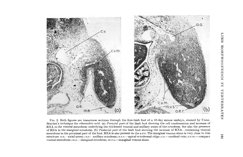
FIG. 2. Both figures are transverse sections through the fore-limb bud of a 10-day mouse embryo, stained by UnnaBrachet's technique for ribonucleic acid, (a) Preaxial part of the limb bud showing the cell condensation and increase of
RNA in the ventral mesoderm underlying the thickened ventral and axillary zones of the ectoderm. See also the presence
of RNA in the marginal ectoderm, (b) Postaxial part of the limb bud showing the increase of RNA—containing ventral
mesoderm in the proximal part of the bud. RNA is also present in the a.e.r. The marginal venous sinus is very close to this
structure, a.a.—axial artery; a.e.—axillary ectoderm; a.e.r.—apical ectodermal ridge; c.v.—cardinal vein; c.v.m.—compact
ventral mesoderm; m.e.—marginal ectoderm; m.v.s.—marginal venous sinus.
g
g
o
«
o
Q
H
H
m
<
H
td
H
x.v.m.
v. (a)·
FIG. 2. Both figures are transverse sections through the fore-limb bud of a 10-day mouse embryo, stained by UnnaBrachet's technique for ribonucleic acid, (a) Preaxial part of the limb bud showing the cell condensation and increase of
RNA in the ventral mesoderm underlying the thickened ventral and axillary zones of the ectoderm. See also the presence
of RNA in the marginal ectoderm, (b) Postaxial part of the limb bud showing the increase of RNA—containing ventral
mesoderm in the proximal part of the bud. RNA is also present in the a.e.r. The marginal venous sinus is very close to this
structure, a.a.—axial artery; a.e.—axillary ectoderm; a.e.r.—apical ectodermal ridge; c.v.—cardinal vein; c.v.m.—compact
ventral mesoderm; m.e.—marginal ectoderm; m.v.s.—marginal venous sinus.
g
g
o
«
o
Q
H
H
m
<
H
td
H