

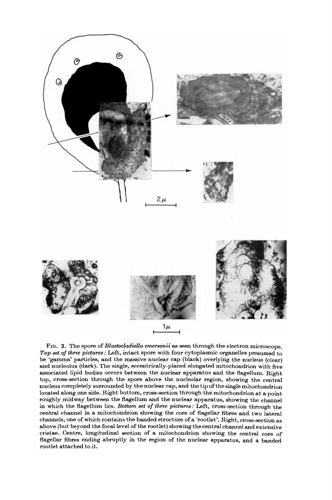
FIG. 3. The spore of Blastocladiella emersonii as seen through the electron microscope.
Top set of three pictures : Left, intact spore with four cytoplasmic organelles presumed to
be 'gamma' particles, and the massive nuclear cap (black) overlying the nucleus (clear)
and nucleolus (dark). The single, eccentrically-placed elongated mitochondrion with five
associated lipid bodies occurs between the nuclear apparatus and the flagellum. Right
top, cross-section through the spore above the nucleolar region, showing the central
nucleus completely surrounded by the nuclear cap, and the tip of the single mitochondrion
located along one side. Right bottom, cross-section through the mitochondrion at a point
roughly midway between the flagellum and the nuclear apparatus, showing the channel
in which the flagellum lies. Bottom set of three pictures : Left, cross-section through the
central channel in a mitochondrion showing the core of flagellar fibres and two lateral
channels, one of which contains the banded structure of a 'rootlet'. Right, cross-section as
above (but beyond the focal level of the rootlet) showing the central channel and extensive
cristae. Centre, longitudinal section of a mitochondrion showing the central core of
flagellar fibres ending abruptly in the region of the nuclear apparatus, and a banded
rootlet attached to it.
Top set of three pictures : Left, intact spore with four cytoplasmic organelles presumed to
be 'gamma' particles, and the massive nuclear cap (black) overlying the nucleus (clear)
and nucleolus (dark). The single, eccentrically-placed elongated mitochondrion with five
associated lipid bodies occurs between the nuclear apparatus and the flagellum. Right
top, cross-section through the spore above the nucleolar region, showing the central
nucleus completely surrounded by the nuclear cap, and the tip of the single mitochondrion
located along one side. Right bottom, cross-section through the mitochondrion at a point
roughly midway between the flagellum and the nuclear apparatus, showing the channel
in which the flagellum lies. Bottom set of three pictures : Left, cross-section through the
central channel in a mitochondrion showing the core of flagellar fibres and two lateral
channels, one of which contains the banded structure of a 'rootlet'. Right, cross-section as
above (but beyond the focal level of the rootlet) showing the central channel and extensive
cristae. Centre, longitudinal section of a mitochondrion showing the central core of
flagellar fibres ending abruptly in the region of the nuclear apparatus, and a banded
rootlet attached to it.