

EGG CORTEX IN MORPHOGENESIS
385
group (Urodela or Anura). The eggs of both groups have, in the
unfertilized state, a
c
zona radiata' i.e. numerous cortical microvilli
embedded in the chorion, and in both cases these microvilli retract and
disappear after fertilization. The difference lies in the presence in the
cortex of the Anura of cortical granules the content of which is expelled
and causes the formation of the perivitelline cavity by uptake of water
from the outside. These cortical granules are not present in the eggs of
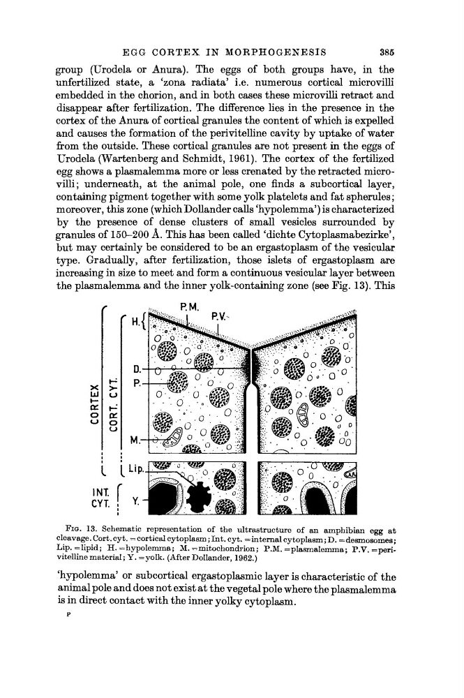
Urodela (Wartenberg and Schmidt, 1961). The cortex of the fertilized
egg shows a plasmalemma more or less crenated by the retracted microvilli; underneath, at the animal pole, one finds a subcortical layer,
containing pigment together with some yolk platelets and fat spherules ;
moreover, this zone (which Dollander calls 'hypolemma') is characterized
by the presence of dense clusters of small vesicles surrounded by
granules of 150-200 Â. This has been called 'dichte Cytoplasmabezirke',
but may certainly be considered to be an ergastoplasm of the vesicular
type. Gradually, after fertilization, those islets of ergastoplasm are
increasing in size to meet and form a continuous vesicular layer between
the plasmalemma and the inner yolk-containing zone (see Fig. 13). This
FIG. 13. Schematic representation of the ultrastructure of an amphibian egg at
cleavage. Cort. cyt. = cortical cytoplasm ; Int. cyt. = internal cytoplasm ; D. = desmosomes ;
Lip. = lipid; H. = hypolemma; M. = mitochondrion; P.M. = plasmalemma; P.V. = perivitelline material ; Y. =yolk. (After Dollander, 1962.)
'hypolemma' or subcortical ergastoplasmic layer is characteristic of the
animal pole and does not exist at the vegetal pole where the plasmalemma
is in direct contact with the inner yolky cytoplasm.
P
385
group (Urodela or Anura). The eggs of both groups have, in the
unfertilized state, a
c
zona radiata' i.e. numerous cortical microvilli
embedded in the chorion, and in both cases these microvilli retract and
disappear after fertilization. The difference lies in the presence in the
cortex of the Anura of cortical granules the content of which is expelled
and causes the formation of the perivitelline cavity by uptake of water
from the outside. These cortical granules are not present in the eggs of
Urodela (Wartenberg and Schmidt, 1961). The cortex of the fertilized
egg shows a plasmalemma more or less crenated by the retracted microvilli; underneath, at the animal pole, one finds a subcortical layer,
containing pigment together with some yolk platelets and fat spherules ;
moreover, this zone (which Dollander calls 'hypolemma') is characterized
by the presence of dense clusters of small vesicles surrounded by
granules of 150-200 Â. This has been called 'dichte Cytoplasmabezirke',
but may certainly be considered to be an ergastoplasm of the vesicular
type. Gradually, after fertilization, those islets of ergastoplasm are
increasing in size to meet and form a continuous vesicular layer between
the plasmalemma and the inner yolk-containing zone (see Fig. 13). This
FIG. 13. Schematic representation of the ultrastructure of an amphibian egg at
cleavage. Cort. cyt. = cortical cytoplasm ; Int. cyt. = internal cytoplasm ; D. = desmosomes ;
Lip. = lipid; H. = hypolemma; M. = mitochondrion; P.M. = plasmalemma; P.V. = perivitelline material ; Y. =yolk. (After Dollander, 1962.)
'hypolemma' or subcortical ergastoplasmic layer is characteristic of the
animal pole and does not exist at the vegetal pole where the plasmalemma
is in direct contact with the inner yolky cytoplasm.
P