

DEVELOPMENT OF GASTROPODS
27
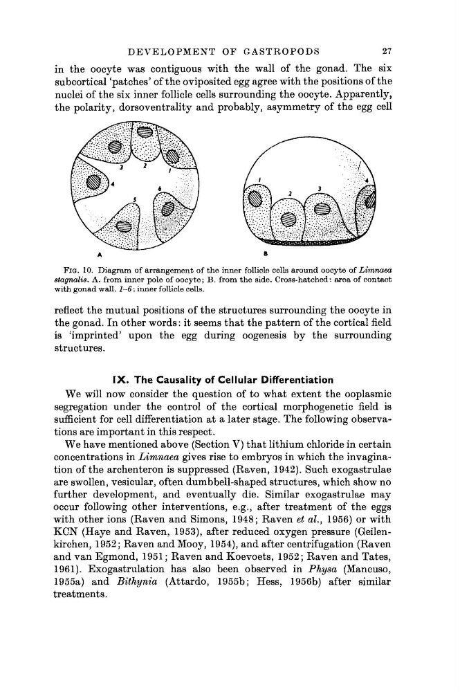
FIG. 10. Diagram of arrangement of the inner follicle cells around oocyte of Limnaea
stagnalis. A. from inner pole of oocyte; B. from the side. Cross-hatched: area of contact
with gonad wall. 1-6 : inner follicle cells.
reflect the mutual positions of the structures surrounding the oocyte in
the gonad. In other words : it seems that the pattern of the cortical field
is 'imprinted' upon the egg during oogenesis by the surrounding
structures.
IX. The Causality of Cellular Differentiation
We will now consider the question of to what extent the ooplasmic
segregation under the control of the cortical morphogenetic field is
sufficient for cell differentiation at a later stage. The following observations are important in this respect.
We have mentioned above (Section V) that lithium chloride in certain
concentrations in Limnaea gives rise to embryos in which the invagination of the archenteron is suppressed (Raven, 1942). Such exogastrulae
are swollen, vesicular, often dumbbell-shaped structures, which show no
further development, and eventually die. Similar exogastrulae may
occur following other interventions, e.g., after treatment of the eggs
with other ions (Raven and Simons, 1948; Raven et al., 1956) or with
KCN (Haye and Raven, 1953), after reduced oxygen pressure (Geilenkirchen, 1952; Raven and Mooy, 1954), and after centrifugation (Raven
and van Egmond, 1951 ; Raven and Koevoets, 1952; Raven and Tates,
1961). Exogastrulation has also been observed in Physa (Mancuso,
1955a) and Bithynia
(Attardo, 1955b; Hess, 1956b) after similar
treatments.
in the oocyte was contiguous with the wall of the gonad. The six
subcortical 'patches' of the oviposited egg agree with the positions of the
nuclei of the six inner follicle cells surrounding the oocyte. Apparently,
the polarity, dorsoventrality and probably, asymmetry of the egg cell
27
FIG. 10. Diagram of arrangement of the inner follicle cells around oocyte of Limnaea
stagnalis. A. from inner pole of oocyte; B. from the side. Cross-hatched: area of contact
with gonad wall. 1-6 : inner follicle cells.
reflect the mutual positions of the structures surrounding the oocyte in
the gonad. In other words : it seems that the pattern of the cortical field
is 'imprinted' upon the egg during oogenesis by the surrounding
structures.
IX. The Causality of Cellular Differentiation
We will now consider the question of to what extent the ooplasmic
segregation under the control of the cortical morphogenetic field is
sufficient for cell differentiation at a later stage. The following observations are important in this respect.
We have mentioned above (Section V) that lithium chloride in certain
concentrations in Limnaea gives rise to embryos in which the invagination of the archenteron is suppressed (Raven, 1942). Such exogastrulae
are swollen, vesicular, often dumbbell-shaped structures, which show no
further development, and eventually die. Similar exogastrulae may
occur following other interventions, e.g., after treatment of the eggs
with other ions (Raven and Simons, 1948; Raven et al., 1956) or with
KCN (Haye and Raven, 1953), after reduced oxygen pressure (Geilenkirchen, 1952; Raven and Mooy, 1954), and after centrifugation (Raven
and van Egmond, 1951 ; Raven and Koevoets, 1952; Raven and Tates,
1961). Exogastrulation has also been observed in Physa (Mancuso,
1955a) and Bithynia
(Attardo, 1955b; Hess, 1956b) after similar
treatments.
in the oocyte was contiguous with the wall of the gonad. The six
subcortical 'patches' of the oviposited egg agree with the positions of the
nuclei of the six inner follicle cells surrounding the oocyte. Apparently,
the polarity, dorsoventrality and probably, asymmetry of the egg cell