

ANIMALIZATION AND VEGETALIZATION
151
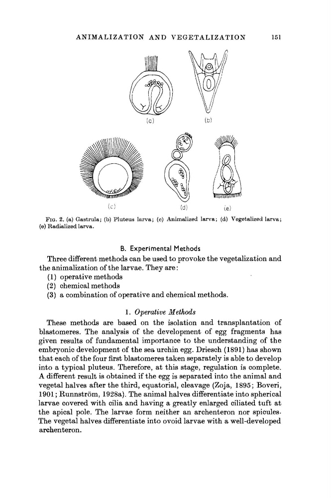
FIG. 2. (a) Gastrula; (b) Pluteus larva; (c) Annualized larva; (d) Vegetalized larva;
(e) Radialized larva.
B. Experimental Methods
Three different methods can be used to provoke the vegetalization and
the animalization of the larvae. They are :
(1) operative methods
(2) chemical methods
(3) a combination of operative and chemical methods.
1. Operative
Methods
These methods are based on the isolation and transplantation of
blastomeres. The analysis of the development of egg fragments has
given results of fundamental importance to the understanding of the
embryonic development of the sea urchin egg. Driesch (1891) has shown
that each of the four first blastomeres taken separately is able to develop
into a typical pluteus. Therefore, at this stage, regulation is complete.
A different result is obtained if the egg is separated into the animal and
vegetal halves after the third, equatorial, cleavage (Zoja, 1895; Boveri,
1901 ; Runnström, 1928a). The animal halves differentiate into spherical
larvae covered with cilia and having a greatly enlarged ciliated tuft at
the apical pole. The larvae form neither an archenteron nor spicules.
The vegetal halves differentiate into ovoid larvae with a well-developed
archenteron.
151
FIG. 2. (a) Gastrula; (b) Pluteus larva; (c) Annualized larva; (d) Vegetalized larva;
(e) Radialized larva.
B. Experimental Methods
Three different methods can be used to provoke the vegetalization and
the animalization of the larvae. They are :
(1) operative methods
(2) chemical methods
(3) a combination of operative and chemical methods.
1. Operative
Methods
These methods are based on the isolation and transplantation of
blastomeres. The analysis of the development of egg fragments has
given results of fundamental importance to the understanding of the
embryonic development of the sea urchin egg. Driesch (1891) has shown
that each of the four first blastomeres taken separately is able to develop
into a typical pluteus. Therefore, at this stage, regulation is complete.
A different result is obtained if the egg is separated into the animal and
vegetal halves after the third, equatorial, cleavage (Zoja, 1895; Boveri,
1901 ; Runnström, 1928a). The animal halves differentiate into spherical
larvae covered with cilia and having a greatly enlarged ciliated tuft at
the apical pole. The larvae form neither an archenteron nor spicules.
The vegetal halves differentiate into ovoid larvae with a well-developed
archenteron.