

ANIMALIZATION AND VEGETALIZATION
149
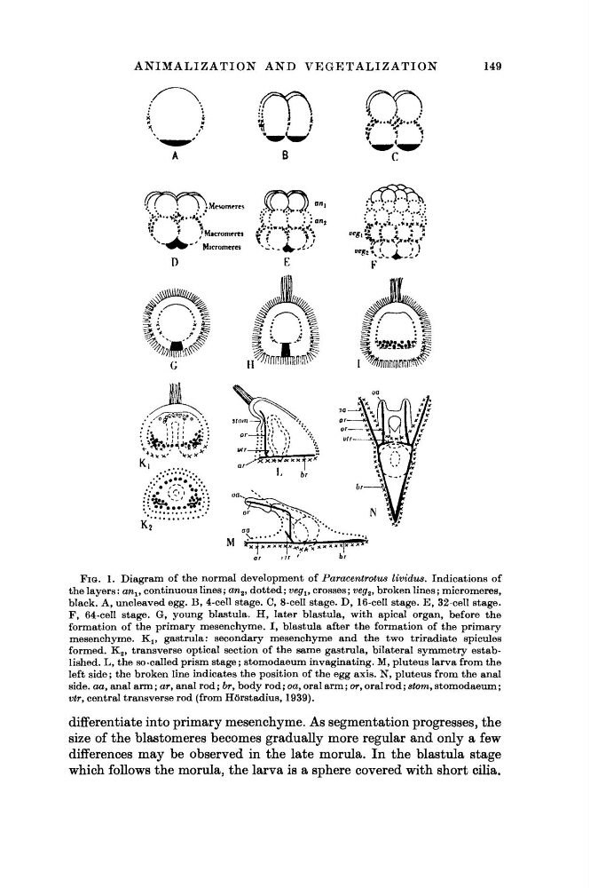
FIG. 1. Diagram of the normal development of Paracentrotus lividus. Indications of
the layers : an lf continuous lines ; an 2 , dotted ; veg x , crosses ; veg 2 , broken lines ; micromeres,
black. A, uncleaved egg. B, 4-cell stage. C, 8-cell stage. D, 16-cell stage. E, 32-cell stage.
F, 64-cell stage. G, young blastula. H, later blastula, with apical organ, before the
formation of the primary mesenchyme. I, blastula after the formation of the primary
mesenchyme. K lf gastrula: secondary mesenchyme and the two triradiate spicules
formed. K 2 , transverse optical section of the same gastrula, bilateral symmetry established. L, the so-called prism stage; stomodaeum invaginating. M, pluteus larva from the
left side ; the broken line indicates the position of the egg axis. N, pluteus from the anal
side, αα, anal arm ; ar, anal rod ; br, body rod ; oa, oral arm ; or, oral rod ; stom, stomodaeum ;
vtr, central transverse rod (from Hörstadius, 1939).
differentiate into primary mesenchyme. As segmentation progresses, the
size of the blastomeres becomes gradually more regular and only a few
differences may be observed in the late morula. In the blastula stage
which follows the morula, the larva is a sphere covered with short cilia.
149
FIG. 1. Diagram of the normal development of Paracentrotus lividus. Indications of
the layers : an lf continuous lines ; an 2 , dotted ; veg x , crosses ; veg 2 , broken lines ; micromeres,
black. A, uncleaved egg. B, 4-cell stage. C, 8-cell stage. D, 16-cell stage. E, 32-cell stage.
F, 64-cell stage. G, young blastula. H, later blastula, with apical organ, before the
formation of the primary mesenchyme. I, blastula after the formation of the primary
mesenchyme. K lf gastrula: secondary mesenchyme and the two triradiate spicules
formed. K 2 , transverse optical section of the same gastrula, bilateral symmetry established. L, the so-called prism stage; stomodaeum invaginating. M, pluteus larva from the
left side ; the broken line indicates the position of the egg axis. N, pluteus from the anal
side, αα, anal arm ; ar, anal rod ; br, body rod ; oa, oral arm ; or, oral rod ; stom, stomodaeum ;
vtr, central transverse rod (from Hörstadius, 1939).
differentiate into primary mesenchyme. As segmentation progresses, the
size of the blastomeres becomes gradually more regular and only a few
differences may be observed in the late morula. In the blastula stage
which follows the morula, the larva is a sphere covered with short cilia.