

2 . M E T H O D S
73
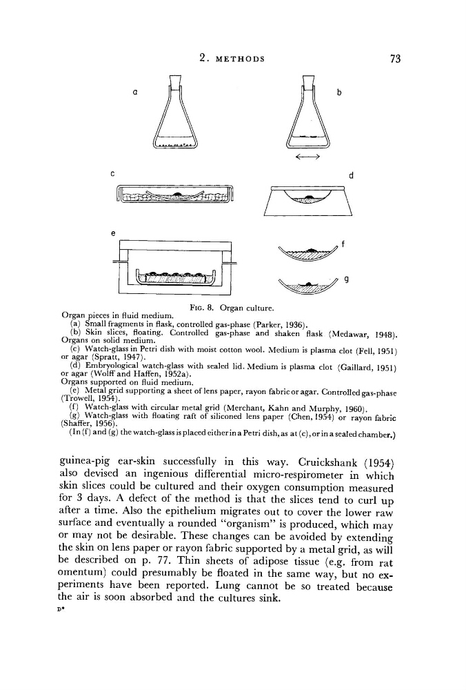
FIG. 8. Organ culture.
Organ pieces in fluid medium.
(a) Small fragments in flask, controlled gas-phase (Parker, 1936).
(b) Skin slices, floating. Controlled gas-phase and shaken flask (Medawar, 1948).
Organs on solid medium.
(c) Watch-glass in Petri dish with moist cotton wool. Medium is plasma clot (Fell, 1951)
or agar (Spratt, 1947).
(d) Embryological watch-glass with sealed lid. Medium is plasma clot (Gaillard, 1951)
or agar (Wolff and Haffen, 1952a).
Organs supported on fluid medium.
(e) Metal grid supporting a sheet of lens paper, rayon fabric or agar. Controlled gas-phase
(Trowell, 1954).
(f) Watch-glass with circular metal grid (Merchant, Kahn and Murphy, 1960).
(g) Watch-glass with floating raft of siliconed lens paper (Chen, 1954) or rayon fabric
(Shaffer, 1956).
(In (f) and (g) the watch-glass is placed either in a Petri dish, as at (c), or in a sealed chamber.)
guinea-pig ear-skin successfully in this way. Cruickshank (1954)
also devised an ingenious differential micro-respirometer in which
skin slices could be cultured and their oxygen consumption measured
for 3 days. A defect of the method is that the slices tend to curl up
after a time. Also the epithelium migrates out to cover the lower raw
surface and eventually a rounded "organism" is produced, which may
or may not be desirable. These changes can be avoided by extending
the skin on lens paper or rayon fabric supported by a metal grid, as will
be described on p. 77. Thin sheets of adipose tissue (e.g. from rat
omentum) could presumably be floated in the same way, but no experiments have been reported. Lung cannot be so treated because
the air is soon absorbed and the cultures sink.
D*
a
c
d
e
f
9
73
FIG. 8. Organ culture.
Organ pieces in fluid medium.
(a) Small fragments in flask, controlled gas-phase (Parker, 1936).
(b) Skin slices, floating. Controlled gas-phase and shaken flask (Medawar, 1948).
Organs on solid medium.
(c) Watch-glass in Petri dish with moist cotton wool. Medium is plasma clot (Fell, 1951)
or agar (Spratt, 1947).
(d) Embryological watch-glass with sealed lid. Medium is plasma clot (Gaillard, 1951)
or agar (Wolff and Haffen, 1952a).
Organs supported on fluid medium.
(e) Metal grid supporting a sheet of lens paper, rayon fabric or agar. Controlled gas-phase
(Trowell, 1954).
(f) Watch-glass with circular metal grid (Merchant, Kahn and Murphy, 1960).
(g) Watch-glass with floating raft of siliconed lens paper (Chen, 1954) or rayon fabric
(Shaffer, 1956).
(In (f) and (g) the watch-glass is placed either in a Petri dish, as at (c), or in a sealed chamber.)
guinea-pig ear-skin successfully in this way. Cruickshank (1954)
also devised an ingenious differential micro-respirometer in which
skin slices could be cultured and their oxygen consumption measured
for 3 days. A defect of the method is that the slices tend to curl up
after a time. Also the epithelium migrates out to cover the lower raw
surface and eventually a rounded "organism" is produced, which may
or may not be desirable. These changes can be avoided by extending
the skin on lens paper or rayon fabric supported by a metal grid, as will
be described on p. 77. Thin sheets of adipose tissue (e.g. from rat
omentum) could presumably be floated in the same way, but no experiments have been reported. Lung cannot be so treated because
the air is soon absorbed and the cultures sink.
D*
a
c
d
e
f
9