

17.
A C T I O N S O F V I T A M I N S A A N D C
675
vitamin, were undertaken to see whether under in vitro conditions the
bones would respond to the vitamin, as this would provide conclusive
evidence for a direct action of the agent on the tissues.
(a) Histological changes in cartilage and bone. These experiments (Fell
and Mellanby, 1952) were made on the cartilaginous rudiments from
6-7-day chick embryos and on late foetal mouse bones; the latter are
well developed with a stout bony shaft, enclosing a marrow cavity, and
large cartilaginous ends.
After 2-3 days' growth in the presence of vitamin A (10 i.u./ml of
medium) the rudiments, both chick and mouse, begin to shrink
(Fig. 16). The cartilage becomes soft, the intercellular partitions
narrower and the matrix loses its metachromasia with such stains as
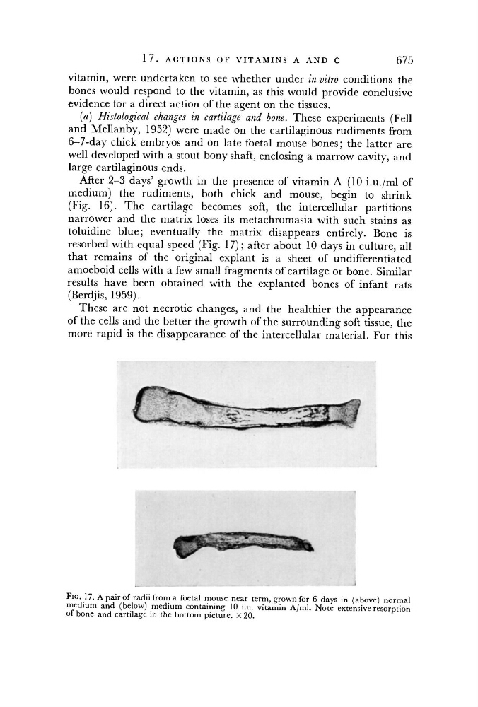
toluidine blue; eventually the matrix disappears entirely. Bone is
resorbed with equal speed (Fig. 17); after about 10 days in culture, all
that remains of the original explant is a sheet of undifferentiated
amoeboid cells with a few small fragments of cartilage or bone. Similar
results have been obtained with the explanted bones of infant rats
(Berdjis, 1959).
These are not necrotic changes, and the healthier the appearance
of the cells and the better the growth of the surrounding soft tissue, the
more rapid is the disappearance of the intercellular material. For this
FIG 17. A pair of radii from a foetal mouse near term, grown for 6 days in (above) normal
medium and (below) medium containing 10 i.u. vitamin A/ml. Note extensive resorption
of bone and cartilage in the bottom picture. X 20.