

17. A C T I O N S OF V I T A M I N S A A N D C
669
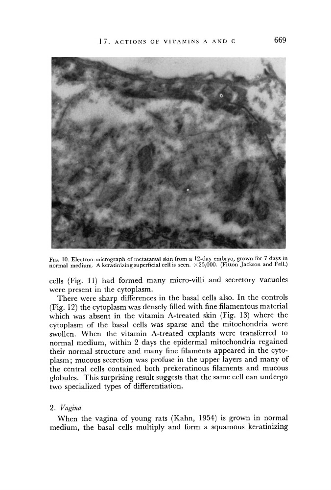
FIG. 10. Electron-micrograph of metatarsal skin from a 12-day embryo, grown for 7 days in
normal medium. A keratinizing superficial cell is seen. X 25,000. (Fitton Jackson and Fell.)
cells (Fig. 11) had formed many micro-villi and secretory vacuoles
were present in the cytoplasm.
There were sharp differences in the basal cells also. In the controls
(Fig. 12) the cytoplasm was densely filled with fine filamentous material
which was absent in the vitamin A-treated skin (Fig. 13) where the
cytoplasm of the basal cells was sparse and the mitochondria were
swollen. When the vitamin A-treated explants were transferred to
normal medium, within 2 days the epidermal mitochondria regained
their normal structure and many fine filaments appeared in the cytoplasm; mucous secretion was profuse in the upper layers and many of
the central cells contained both prekeratinous filaments and mucous
globules. This surprising result suggests that the same cell can undergo
two specialized types of differentiation.
2. Vagina
When the vagina of young rats (Kahn, 1954) is grown in normal
medium, the basal cells multiply and form a squamous keratinizing