

598
I L S E
L A S N I T Z K I
vation revealed a disturbance of cartilage differentiation. The zone
of flattened cells was either poorly defined or entirely absent, so that
the epiphysis merged imperceptibly with the shaft. The diaphyseal
cells never developed beyond their original stage of hypertrophy; they
were smaller and their capsules ill-defined. Staining with toluidine blue
showed that the matrix stained less intensely metachromatically than
in the controls. The epiphysis, although abnormally large, was histologically almost normal. The amount of periosteal bone seemed to
depend on the stage of histogenesis at the time of explantation. Rudiments in which chondroblastic hypertrophy was well advanced such
as in most femora, tibiae and humeri, produced more bone, while
in all radii and ulnae, which were less advanced, periosteal ossification
was inferior to that of the controls. Some rudiments were completely
devoid of bone and remained a uniform mass of cartilage.
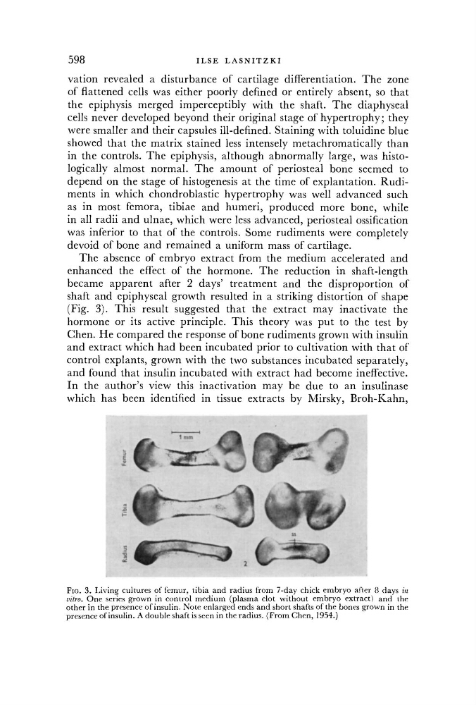
The absence of embryo extract from the medium accelerated and
enhanced the effect of the hormone. The reduction in shaft-length
became apparent after 2 days' treatment and the disproportion of
shaft and epiphyseal growth resulted in a striking distortion of shape
(Fig. 3). This result suggested that the extract may inactivate the
hormone or its active principle. This theory was put to the test by
Chen. He compared the response of bone rudiments grown with insulin
and extract which had been incubated prior to cultivation with that of
control explants, grown with the two substances incubated separately,
and found that insulin incubated with extract had become ineffective.
In the author's view this inactivation may be due to an insulinase
which has been identified in tissue extracts by Mirsky, Broh-Kahn,
FIG. 3. Living cultures of femur, tibia and radius from 7-day chick embryo after 8 days in
vitro. One series grown in control medium (plasma clot without embryo extract) and the
other in the presence of insulin. Note enlarged ends and short shafts of the bones grown in the
presence of insulin. A double shaft is seen in the radius. (From Chen, 1954.)
I L S E
L A S N I T Z K I
vation revealed a disturbance of cartilage differentiation. The zone
of flattened cells was either poorly defined or entirely absent, so that
the epiphysis merged imperceptibly with the shaft. The diaphyseal
cells never developed beyond their original stage of hypertrophy; they
were smaller and their capsules ill-defined. Staining with toluidine blue
showed that the matrix stained less intensely metachromatically than
in the controls. The epiphysis, although abnormally large, was histologically almost normal. The amount of periosteal bone seemed to
depend on the stage of histogenesis at the time of explantation. Rudiments in which chondroblastic hypertrophy was well advanced such
as in most femora, tibiae and humeri, produced more bone, while
in all radii and ulnae, which were less advanced, periosteal ossification
was inferior to that of the controls. Some rudiments were completely
devoid of bone and remained a uniform mass of cartilage.
The absence of embryo extract from the medium accelerated and
enhanced the effect of the hormone. The reduction in shaft-length
became apparent after 2 days' treatment and the disproportion of
shaft and epiphyseal growth resulted in a striking distortion of shape
(Fig. 3). This result suggested that the extract may inactivate the
hormone or its active principle. This theory was put to the test by
Chen. He compared the response of bone rudiments grown with insulin
and extract which had been incubated prior to cultivation with that of
control explants, grown with the two substances incubated separately,
and found that insulin incubated with extract had become ineffective.
In the author's view this inactivation may be due to an insulinase
which has been identified in tissue extracts by Mirsky, Broh-Kahn,
FIG. 3. Living cultures of femur, tibia and radius from 7-day chick embryo after 8 days in
vitro. One series grown in control medium (plasma clot without embryo extract) and the
other in the presence of insulin. Note enlarged ends and short shafts of the bones grown in the
presence of insulin. A double shaft is seen in the radius. (From Chen, 1954.)