

512
A . A . M O S C O N A
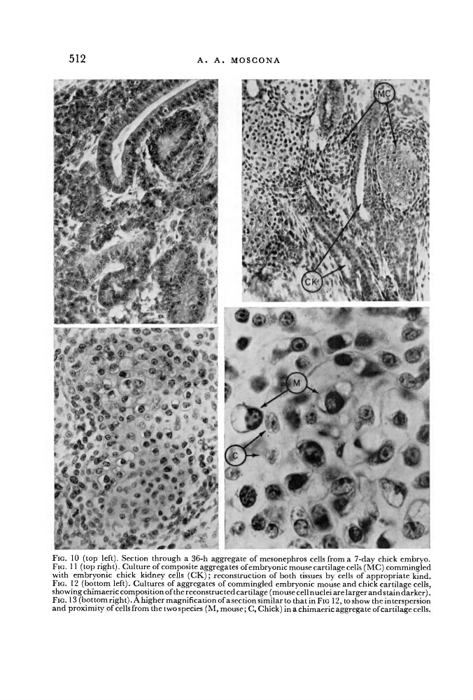
FIG. 10 (top left). Section through a 36-h aggregate of mesonephros cells from a 7-day chick embryo.
FIG. 11 (top right). Culture of composite aggregates of embryonic mouse cartilage cells (MC) commingled
with embryonic chick kidney cells ( C K ) ; reconstruction of both tissues by cells of appropriate kind.
FIG. 12 (bottom left). Cultures of aggregates of commingled embryonic mouse and chick cartilage cells,
showing chimaeric composition of the reconstructed cartilage (mouse cell nuclei are larger and stain darker).
FIG. 13 (bottom right). A higher magnification of a section similar to that in FIG 12, to show the interspersion
and proximity of cells from the two species ( M , mouse; C, Chick) in a chimaeric aggregate of cartilage cells.
A . A . M O S C O N A
FIG. 10 (top left). Section through a 36-h aggregate of mesonephros cells from a 7-day chick embryo.
FIG. 11 (top right). Culture of composite aggregates of embryonic mouse cartilage cells (MC) commingled
with embryonic chick kidney cells ( C K ) ; reconstruction of both tissues by cells of appropriate kind.
FIG. 12 (bottom left). Cultures of aggregates of commingled embryonic mouse and chick cartilage cells,
showing chimaeric composition of the reconstructed cartilage (mouse cell nuclei are larger and stain darker).
FIG. 13 (bottom right). A higher magnification of a section similar to that in FIG 12, to show the interspersion
and proximity of cells from the two species ( M , mouse; C, Chick) in a chimaeric aggregate of cartilage cells.