

6. CELL D I V I S I O N
205
In vertebrate cultures, prophase has an imprecise beginning. It starts
perhaps by a rotation of the nucleus (Commandon, de Fonbrunne and
Jolly, 1934; Hughes, 1952) happening too early to be caught often by
the cine camera. The chromosomes as seen with phase contrast become
more and more distinct but remain inside the nuclear envelope.
Prophase ends by the sudden fading of the nucleolus and disappearance
of the nuclear membrane. Prometaphase is a variable period. The
chromosomes spread slowly out of the former nuclear area and attach
themselves one by one to the spindle. Characteristic to and fro movements of the chromosomes continue until they are all attached. Then,
suddenly, the group freezes into an equatorial plate. This is the
metaphase proper, usually short and sometimes coinciding with the
beginning of peripheral cytoplasmic ''bubbling". The onset of anaphase,
when the chromosomes begin to draw apart, is a sharply defined event,
perhaps the most dramatic moment of the division. Telophase is considered to begin with the constriction of the cytoplasm in the area
where the equatorial plate stood a few minutes before. It is prolonged
into a reconstruction phase (so-called for lack of a better name) which
continues after the reappearance of the nucleolus and the nuclear
membrane and even after the separation of the two daughter cells. The
nucleus is then still easily recognized by its granular aspect, due to the
long time taken by chromosomes to fade into intermitotic chromatin.
2. Duration
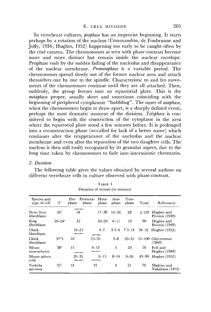
The following table gives the values obtained by several authors on
different vertebrate cells in culture observed with phase-contrast.
TABLE I
Duration of mitosis (in minutes)
Species and
type of cell
T°
Pro- Prometa- Metaphase
phase
phase
Anaphase
Telophase
Total
References
Newt liver
26°
17-38
14-26
28
± 120 Hughes and
fibroblasts
Preston (1949)
Frog
20-24°
32
20-29
6-11
16
90
Hughes and
fibroblasts
Preston (1949)
Chick
19-25
4-7
3-5-6 7-5-14 34-52 Hughes (1952)
fibroblasts
' — • — '
Hughes (1952)
Chick
37°5
10
25-35
3-8
20-35 55-100 Chevremont
fibroblasts
(1960)
Mouse
38°
15
6-13
5
20
59
Fell and
mesenchyme
' — • —
Hughes (1949)
Mouse spleen
20-35
6-15
8-14
9-26
43-90 Hughes (1952)
cells
• — • — •
• — " — •
Yoshida
35°
14
31
4
21
70
Makino and
sarcoma
Nakahara(1953)
205
In vertebrate cultures, prophase has an imprecise beginning. It starts
perhaps by a rotation of the nucleus (Commandon, de Fonbrunne and
Jolly, 1934; Hughes, 1952) happening too early to be caught often by
the cine camera. The chromosomes as seen with phase contrast become
more and more distinct but remain inside the nuclear envelope.
Prophase ends by the sudden fading of the nucleolus and disappearance
of the nuclear membrane. Prometaphase is a variable period. The
chromosomes spread slowly out of the former nuclear area and attach
themselves one by one to the spindle. Characteristic to and fro movements of the chromosomes continue until they are all attached. Then,
suddenly, the group freezes into an equatorial plate. This is the
metaphase proper, usually short and sometimes coinciding with the
beginning of peripheral cytoplasmic ''bubbling". The onset of anaphase,
when the chromosomes begin to draw apart, is a sharply defined event,
perhaps the most dramatic moment of the division. Telophase is considered to begin with the constriction of the cytoplasm in the area
where the equatorial plate stood a few minutes before. It is prolonged
into a reconstruction phase (so-called for lack of a better name) which
continues after the reappearance of the nucleolus and the nuclear
membrane and even after the separation of the two daughter cells. The
nucleus is then still easily recognized by its granular aspect, due to the
long time taken by chromosomes to fade into intermitotic chromatin.
2. Duration
The following table gives the values obtained by several authors on
different vertebrate cells in culture observed with phase-contrast.
TABLE I
Duration of mitosis (in minutes)
Species and
type of cell
T°
Pro- Prometa- Metaphase
phase
phase
Anaphase
Telophase
Total
References
Newt liver
26°
17-38
14-26
28
± 120 Hughes and
fibroblasts
Preston (1949)
Frog
20-24°
32
20-29
6-11
16
90
Hughes and
fibroblasts
Preston (1949)
Chick
19-25
4-7
3-5-6 7-5-14 34-52 Hughes (1952)
fibroblasts
' — • — '
Hughes (1952)
Chick
37°5
10
25-35
3-8
20-35 55-100 Chevremont
fibroblasts
(1960)
Mouse
38°
15
6-13
5
20
59
Fell and
mesenchyme
' — • —
Hughes (1949)
Mouse spleen
20-35
6-15
8-14
9-26
43-90 Hughes (1952)
cells
• — • — •
• — " — •
Yoshida
35°
14
31
4
21
70
Makino and
sarcoma
Nakahara(1953)