

370
w. B. REDMOND et al.
Prototype strains for each subtype of M . tuberculosis have been selected
and are available. These will be distributed with the phages.
Plates should be checked daily beginning 24 h after spotting and
records kept of lysis for each dilution for 10 days. The final results to be
reported should be the maximal lysis of each phage for each bacterial
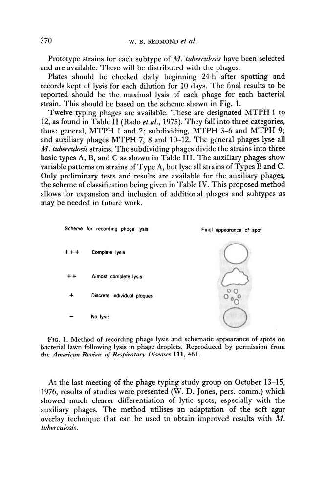
strain. This should be based on the scheme shown in Fig. 1.
Twelve typing phages are available. These are designated M T P H 1 to
12, as found in Table I1 (Rado et al., 1975). They fall into three categories,
thus: general, M T P H 1 and 2; subdividing, M T P H 3-6 and M T P H 9;
and auxiliary phages M T P H 7, 8 and 10-12. The general phages lyse all
M . tuberculosis strains. The subdividing phages divide the strains into three
basic types A, B, and C as shown in Table 111. The auxiliary phages show
variable patterns on strains of Type A, but lyse all strains of Types B and C.
Only preliminary tests and results are available for the auxiliary phages,
the scheme of classification being given in Table IV. This proposed method
allows for expansion and inclusion of additional phages and subtypes as
may be needed in future work.
Scheme for recording phage lysis
Final appearance of spot
+ + + Complete lysis
++ Almost complete lysis
+
Discrete individual ploques
-
No lysis
FIG. 1 . Method of recording phage lysis and schematic appearance of spots on
bacterial lawn following lysis in phage droplets. Reproduced by permission from
the American Review of Respiratory Diseases 111, 461.
At the last meeting of the phage typing study group on October 13-15,
1976, results of studies were presented (W. D. Jones, pers. comm.) which
showed much clearer differentiation of lytic spots, especially with the
auxiliary phages. The method utilises an adaptation of the soft agar
overlay technique that can be used to obtain improved results with M.
tuberculosis.
w. B. REDMOND et al.
Prototype strains for each subtype of M . tuberculosis have been selected
and are available. These will be distributed with the phages.
Plates should be checked daily beginning 24 h after spotting and
records kept of lysis for each dilution for 10 days. The final results to be
reported should be the maximal lysis of each phage for each bacterial
strain. This should be based on the scheme shown in Fig. 1.
Twelve typing phages are available. These are designated M T P H 1 to
12, as found in Table I1 (Rado et al., 1975). They fall into three categories,
thus: general, M T P H 1 and 2; subdividing, M T P H 3-6 and M T P H 9;
and auxiliary phages M T P H 7, 8 and 10-12. The general phages lyse all
M . tuberculosis strains. The subdividing phages divide the strains into three
basic types A, B, and C as shown in Table 111. The auxiliary phages show
variable patterns on strains of Type A, but lyse all strains of Types B and C.
Only preliminary tests and results are available for the auxiliary phages,
the scheme of classification being given in Table IV. This proposed method
allows for expansion and inclusion of additional phages and subtypes as
may be needed in future work.
Scheme for recording phage lysis
Final appearance of spot
+ + + Complete lysis
++ Almost complete lysis
+
Discrete individual ploques
-
No lysis
FIG. 1 . Method of recording phage lysis and schematic appearance of spots on
bacterial lawn following lysis in phage droplets. Reproduced by permission from
the American Review of Respiratory Diseases 111, 461.
At the last meeting of the phage typing study group on October 13-15,
1976, results of studies were presented (W. D. Jones, pers. comm.) which
showed much clearer differentiation of lytic spots, especially with the
auxiliary phages. The method utilises an adaptation of the soft agar
overlay technique that can be used to obtain improved results with M.
tuberculosis.