

26
2. TISSUE EXPOSURE
making the perfusion, and we have tried to build up blood carbon
dioxide levels close to the lethal point before beginning perfusion. None
of these methods made possible a satisfactory general perfusion. It is
reasonable to think that similar experiments must have been attempted
in numerous other laboratories, and these too must have failed for the
literature is singularly silent about success with this method with the
notable exception of the work of Palay et al. (1962). The latter authors
achieved success by extremely careful attention to detail.
The principal technique of perfusing osmium tetroxide fixative described by Palay and his co-workers (1962) is applicable to rats and, more
specifically, their central nervous system. A variant technique is described
for the goldfish.
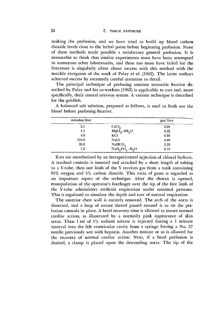
A balanced salt solution, prepared as follows, is used to flush out the
blood before perfusing
fixative.
mmoles/liter
gm/liter
2.5
CaCl2
0.28
1.1
MgCl26H20
0.22
4.0
KC1
0.30
116.0
NaCl
6.80
26.0
NaHC03
2.20
1.2
NaH2P04.H20
0.16
Rats are anesthetized by an intraperitoneal injection of chloral hydrate.
A tracheal cannula is inserted and attached by a short length of tubing
to a Y-tube, then one limb of the Y receives gas from a tank containing
95% oxygen and 5% carbon dioxide. This ratio of gases is regarded as
an important aspect of the technique. After the thorax is opened,
manipulation of the operator's forefinger over the tip of the free limb of
the Y-tube administers artificial respiration under minimal pressure.
This is regulated to simulate the depth and rate of normal respiration.
The anterior chest wall is entirely removed. The arch of the aorta is
dissected, and a loop of suture thread passed around it to tie the perfusion cannula in place. A brief recovery time is allowed to insure normal
cardiac action, as illustrated by a normally pink appearance of skin
areas. Then 1 ml of 1% sodium nitrate is injected during a 1 minute
interval into the left ventricular cavity from a syringe having a No. 27
needle previously wet with heparin. Another minute or so is allowed for
the recovery of normal cardiac action. Next, if a head perfusion is
desired, a clamp is placed upon the descending aorta. The tip of the
2. TISSUE EXPOSURE
making the perfusion, and we have tried to build up blood carbon
dioxide levels close to the lethal point before beginning perfusion. None
of these methods made possible a satisfactory general perfusion. It is
reasonable to think that similar experiments must have been attempted
in numerous other laboratories, and these too must have failed for the
literature is singularly silent about success with this method with the
notable exception of the work of Palay et al. (1962). The latter authors
achieved success by extremely careful attention to detail.
The principal technique of perfusing osmium tetroxide fixative described by Palay and his co-workers (1962) is applicable to rats and, more
specifically, their central nervous system. A variant technique is described
for the goldfish.
A balanced salt solution, prepared as follows, is used to flush out the
blood before perfusing
fixative.
mmoles/liter
gm/liter
2.5
CaCl2
0.28
1.1
MgCl26H20
0.22
4.0
KC1
0.30
116.0
NaCl
6.80
26.0
NaHC03
2.20
1.2
NaH2P04.H20
0.16
Rats are anesthetized by an intraperitoneal injection of chloral hydrate.
A tracheal cannula is inserted and attached by a short length of tubing
to a Y-tube, then one limb of the Y receives gas from a tank containing
95% oxygen and 5% carbon dioxide. This ratio of gases is regarded as
an important aspect of the technique. After the thorax is opened,
manipulation of the operator's forefinger over the tip of the free limb of
the Y-tube administers artificial respiration under minimal pressure.
This is regulated to simulate the depth and rate of normal respiration.
The anterior chest wall is entirely removed. The arch of the aorta is
dissected, and a loop of suture thread passed around it to tie the perfusion cannula in place. A brief recovery time is allowed to insure normal
cardiac action, as illustrated by a normally pink appearance of skin
areas. Then 1 ml of 1% sodium nitrate is injected during a 1 minute
interval into the left ventricular cavity from a syringe having a No. 27
needle previously wet with heparin. Another minute or so is allowed for
the recovery of normal cardiac action. Next, if a head perfusion is
desired, a clamp is placed upon the descending aorta. The tip of the