

Résultats et discussion
42
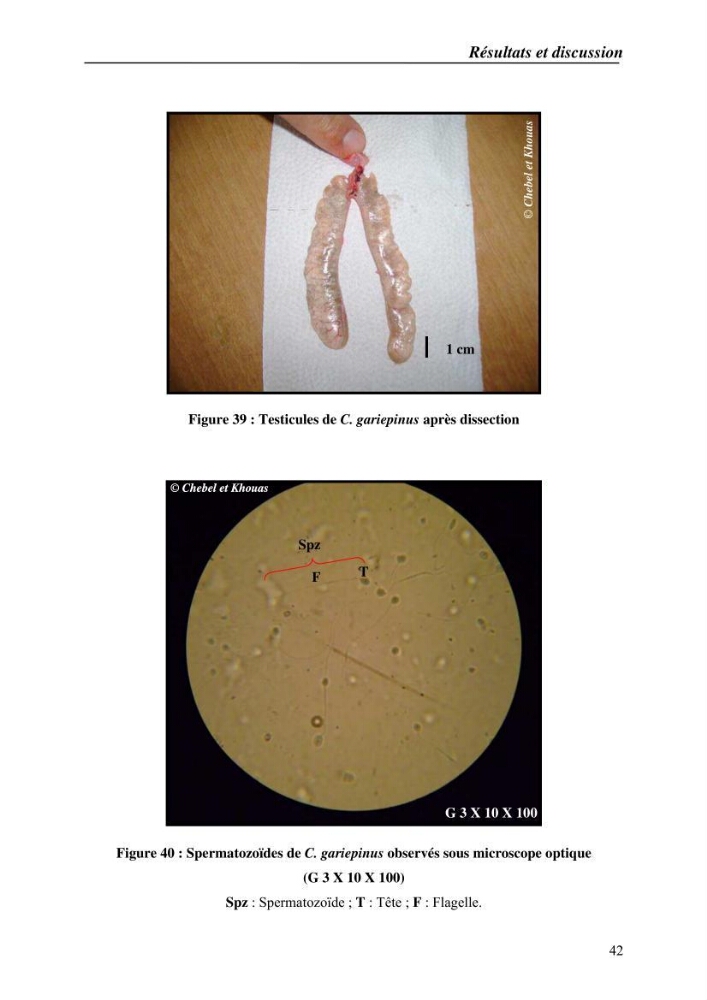
Figure 39 : Testicules de C. gariepinus après dissection
Figure 40 : Spermatozoïdes de C. gariepinus observés sous microscope optique
(G 3 X 10 X 100)
Spz : Spermatozoïde ; T : Tête ; F : Flagelle.
T
F
Spz
G 3 X 10 X 100
© Chebel et Khouas
1 cm
© Chebel et Khouas
© Chebel et Khouas
42
Figure 39 : Testicules de C. gariepinus après dissection
Figure 40 : Spermatozoïdes de C. gariepinus observés sous microscope optique
(G 3 X 10 X 100)
Spz : Spermatozoïde ; T : Tête ; F : Flagelle.
T
F
Spz
G 3 X 10 X 100
© Chebel et Khouas
1 cm
© Chebel et Khouas
© Chebel et Khouas